

 PDF
PDF ePub
ePub Citation
 Print
Print


INTRODUCTION
Tubulocystic renal carcinoma (TCRC) is a rare subtype of renal cell carcinoma that has not yet been included in the World Health Organization (WHO) 2004 classification of renal tumors (1). This tumor entity occurs predominantly in males, and has a specific macroscopic spongy appearance and microscopic characteristics presenting as cysts lined by hobnail cells separated by a thin fibrotic stroma (23).
To date, few published case reports exist on this rare tumor entity. Since most of the previously reported articles have been published in the pathologic literature, little attention has been paid to the radiological features of TCRC.
Herein, we present a case of a 22-year-old woman with TCRC which metastasized to the parasymphyseal pubic bone. We describe the imaging findings of computed tomography (CT), ultrasonography (US), and 18-fluoro-2-deoxy-D-glucose positron emission tomography/computed tomography (18F-FDG PET/CT) with histopathologic correlation.
CASE REPORT
A 22-year-old woman with an incidentally discovered renal mass on check-up abdominal US in an outside hospital was admitted to our hospital for further evaluation and management. The general physical examination, laboratory tests, and urine analysis were normal.
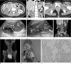
Contrast-enhanced abdomen CT showed a 6.4 cm sized well-defined multilocular cystic mass with enhancing septa and solid portions in the lower pole of the right kidney (Fig. 1A, B). Multifocal curvilinear calcifications and inner septa were seen within the mass. This mass can be classified as a cystic lesion of Bosniak category IV. Also, an osteolytic lesion with soft tissue formation was detected in the left parasymphyseal pubic bone, suggesting pubic bone metastasis (Fig. 1C). Abdominal gray-scale and color Doppler US scan were performed to evaluate a small low attenuation lesion of the liver that was detected on CT. US scan also revealed a lobulating multiseptated heterogeneous cystic mass with calcifications and solid portions which show some inner vascularity in the lower pole of the right kidney (Fig. 1D, E). 18F-FDG PET/CT showed peripheral intense FDG uptake (SUVmax 7.5) with central photon defect in the right renal mass, and also showed another intense FDG uptake (SUVmax 7.2) in the left parasymphyseal pubic bone (Fig. 1F, G).
The patient underwent a radical nephrectomy with dissection of the lymph nodes, and was subsequently treated with a wide marginal excision and allograft for pubic bone metastasis. On pathologic report, the main mass in the right kidney was a well circumscribed, multilocular cystic and solid mass with a spongy appearance, measuring 11.3 × 10.0 × 6.5 cm (Fig. 1H). Microscopic examination revealed that the tumor was composed of variable-sized cysts and well-formed tubules. The epithelial cells had a cuboidal appearance with abundant eosinophilic cytoplasm and prominent nucleoli (Fig. 1I). The cyst lining cells had a hobnail appearance. The tumor cells were positive for epithelial membrane antigen (EMA), CD10 and vimentin, and negative for cytokeratin (CK) 7 and E-cadherin. As a result of these findings, the final histopathologic diagnosis was tubulocystic carcinoma of the kidney. According to the seventh edition of the American Joint Committee on Cancer (AJCC) cancer staging manual, which is based on the extent of the tumor (T), the extent of spread to the lymph nodes (N), and the presence of distant metastasis (M), the right kidney tumor was pT2N0M1.
After the surgery, the patient was treated with immunotherapy, consisting of interleukin-2 (IL-2) at 10 million international units (MIU)/day and interferon-alpha at 6 MIU/day for four weeks. Then, the patient presented with headache and a gadolinium-enhanced brain magnetic resonance imaging revealed a 1.8 × 1.6 cm sized expansile mass at the left petrous bone, just anterior and inferior to the left internal auditory canal. This mass showed low signal intensity (SI) on T1 weighted images (WI), high SI on T2 WI and homogeneous enhancement. Additional temporal bone CT and 18F-FDG PET/CT showed multiple bone metastases in the left temporal bone and the fourth and sixth cervical vertebrae, which were pathologically confirmed as metastatic lesions by following the percutaneous biopsy of the sixth cervical vertebra. Due to disease progression, the patient was treated with 13 cycles of radiotherapy for metastasis at the left temporal bone and the fourth and sixth cervical vertebrae, as well as targeted chemotherapy of sunitinib malate at 50 mg/day for two weeks with one week of rest. Five months after chemotherapy, the patient remained well without signs of any further progression of the metastatic disease.
DISCUSSION
TCRC is a rare renal neoplasm and recently entitled entity that was first described in 1997 by MacLennan et al. (4) as a "low-grade collecting duct carcinoma of the kidney", because at that time, it was thought to be collecting duct origin. However, Amin et al. (5) later renamed this tumor as "tubulocystic carcinoma of the kidney", by showing a distinctive immunostaining that differs from classical collecting duct carcinoma.
Grossly, TCRC is a well-circumscribed tumor that often exhibits a cystic component which may present as radiological classification of Bosniak III or IV. Microscopically, it is composed of tubules and cysts lined by a single layer of tumor cells with eosinophilic cytoplasm and prominent nucleoli, and separated by fibrotic septa (23). Immunohistochemical studies show variable marker positivity with cytokeratins CK7, CK8, CK18, CK19, CD10, and sometimes with E-cadherin (567).
TCRC has a tendency to be seen predominantly in male patients with a wide age range, mostly in the fifth and sixth decade, and usually shows no symptoms. Moreover, most TCRCs have taken an indolent course (4567). In a study on 13 cases of TCRC, seven patients had tumors with pT1, four had with pT2, and two had pT3 (4). Amin et al. (5) reported 31 cases of TCRC, in which 24 patients had tumors with pT1, four patients had with pT2, and three had pT3 at presentation. Follow-up studies in 22 patients showed that one patient had bone metastases, and one patient had bone and liver metastases. As seen above, most TCRCs have taken an indolent course, but some reports shows aggressive metastasis observed in the liver and bone (5). Therefore, TCRC has a low but definite possible potential for metastasis. However, because of the rarity of this tumor, long-term follow-up information of a larger number of cases is necessary for a better prediction of prognosis. Unusually, our patient was a young woman and initially had single bone metastasis without demonstrable lymph node metastasis. However, despite significant surgical and medical treatment, she had an aggressive prognosis presenting as multiple bone metastases.
Since TCRC frequently has cystic components, this tumor entity is often radiologically described as a cystic lesion of Bosniak category III or IV (46). In a recent study of five cases of TCRC, there were two Bosniak III cases, one IV case and two solid tumors (8). Therefore, making a radiological differential diagnosis TCRC from other benign or malignant renal cystic lesions, such as multiloculated clear cell renal cell carcinoma, cystic nephroma, mixed epithelial and stromal tumor and cystic oncocytoma, may be a challenging problem. In our case, TCRC was also classified as a cystic lesion of Bosniak category IV on contrast-enhanced CT images.
In summary, we report a case of a rare aggressive TCRC on the kidney with multiple bone metastases in a young woman that was confirmed on pathology. This tumor presented as a cystic lesion of Bosniak category IV on CT scan with intense FDG uptake on 18F-FDG PET/CT.
 XML Download