

 PDF
PDF ePub
ePub Citation
 Print
Print


INTRODUCTION
Tracheobronchial disruption is an uncommon injury associated with blunt chest trauma (1). According to several articles dealing with radiologic findings of tracheobronchial rupture after blunt chest trauma (2-4), secondary findings of an air leak (pneumomediastinum, subcutaneous emphysema, and persistent pneumothorax despite suction drainage) can be critical hints for the diagnosis of a tracheobronchial rupture. However, these findings are nonspecific in polytraumatized patients, sometimes making an early diagnosis difficult (5,6).
We report CT features and pathologic findings of two pediatric cases in which a bronchial injury was unnoticed initially but was diagnosed later by appearance of delayed bronchostenosis with distal atelectasis after blunt chest trauma in recent motor vehicle accidents.
CASE REPORTS
Case 1
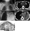
A 7-yr-old boy was admitted to our hospital after a motor vehicle-pedestrian accident. Initial chest radiograph and chest CT scan obtained using a helical CT scanner (HiSpeed Advantage; General Electric Medical Systems, Milwaukee, WI, U.S.A.) on the first hospital day showed extensive pneumomediastinum associated with massive subcutaneous emphysema in the chest wall (Fig. 1A, B). Brain CT scan showed subdural and intracerebral hemorrhage with a skull fracture. Conservative management was done in the intensive care unit.
Ten days later, follow-up anteroposterior chest radiograph showed total collapse of the left lung (Fig. 1C), which thereafter persisted for five days. Contrast-enhanced chest CT scan, which was performed in order to find out the cause of total collapse, revealed airway obliteration at the mid-portion of the left main bronchus with distal collapse (Fig. 1D). The previously noted pneumomediastinum and subcutaneous emphysema were no longer seen.
Total collapse of the left lung persisted at daily routine chest radiographs for six days more, and a bronchoscopic examination was performed, which also showed near-complete obstruction of the left main bronchus. The next day (the 22nd hospital day) we performed bronchial segmental resection and end-to-end anastomosis. At surgery, approximately 5-mm-long, stenotic segment was found at the mid-portion of the left main bronchus.
Pathologic specimen obtained from bronchial segmental resection showed obliteration of the bronchial lumen by fibrous scar and granulation tissue (Fig. 1E), which seemed to be secondary to recent transmural injury of the bronchus. The left lung was fully re-expanded after surgery, and the recovery was uneventful. Follow-up brain CT obtained on the 38th hospital day showed decreased amount of subdural hemorrhage and a cystic change of intracerebral hemorrhage.
Case 2
A 2-yr-old boy was admitted to our hospital after a motor vehicle-pedestrian accident. Initial chest CT scan obtained on the first hospital day showed bilateral hemopneumothoraces, pneumomediastinum, and subcutaneous emphysema in the chest wall with multiple rib fractures. A large consolidation containing multiple cavities was also noted in the right upper lobe, suggestive of traumatic lung cysts. Conservative management including bilateral chest tube placement was done in the intensive care unit.
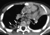
Ten days later, a follow-up anteroposterior chest radiograph showed atelectasis of the left upper lobe, which thereafter persisted for five days at daily routine chest radiographs. Contrast-enhanced chest CT scan, which was performed in order to find out the cause of lobar atelectasis, revealed obliteration of the proximal portion of the left upper lobe bronchus (Fig. 2). The previous bilateral hemopneumothoraces, pneumomediastinum, and subcutaneous emphysema were no longer seen, and the extent of the cavitary consolidation in the right upper lobe has decreased. Since left upper lobe atelectasis persisted for eight days more, a bronchoscopic examination was performed, which showed near-complete obstruction of the left upper lobar bronchus.
Four days after the bronchoscopic examination (the 27th hospital day), bronchial segmental resection and end-to-end anastomosis of the left upper lobar bronchus was performed. At surgery, approximately 5-mm-long, stenotic segment was found at the proximal portion of the left upper lobe bronchus, showing complete luminal obstruction by fibrotic change.
Pathologic specimen obtained from bronchial segmental resection showed obliteration of the bronchial lumen by dense fibrous overgrowth and granulation tissue, which was identical to the pathologic finding of Case 1. The previously atelectatic left upper lobe was fully re-expanded after surgery, and the recovery was uneventful.
DISCUSSION
In most cases, a tracheobronchial rupture is suspected radiologically in front of pneumomediastinum, cervical and thoracic subcutaneous emphysema, and persistent pneumothorax and bronchopleural air leak despite chest tube placement (2). A bronchoscopic examination can confirm the presence and location of the airway rupture, which is reconfirmed and repaired by surgery. Although chest CT scan sometimes visualizes directly the presence of tracheobronchial rupture, it can generally show only secondary findings of an air leak (2).
Secondary findings of an air leak are consequences of free communication between the site of the tracheobronchial disruption and the pleural cavity, which results in a large persistent pneumothorax despite tube thoracotomy (6). If the bronchial transection is incomplete and there is little communication between the proximal transected bronchus and the mediastinum and/or the pleural space, a once-developed pneumomediastinum or pneumothorax will subside well either spontaneously or after chest tube placement. In this circumstance, the tracheobronchial injury will not be noticed radiologically until delayed atelectasis resulting from fibrotic bronchostenosis appears as in our cases (6). In our cases, because the initial pneumomediastinum and subcutaneous emphysema and/or pneumothoraces subsided well either spontaneously or after chest tube placement within 15 days with the vital signs of the patients being stable, the bronchial injury remained unnoticed over a period of two weeks.
On review of literature regarding bronchial rupture after blunt chest trauma, one of two patients reported by Epelman et al. (7) was similar to our cases, in whom a bronchial injury was identified as delayed atelectasis of the left lower lobe with bronchial obliteration at CT ten days after blunt chest trauma. Ozcelik et al. (8) reported a case with combined right main bronchial disruption and chylothorax manifesting as a consolidated right lung and a small pleural effusion that were diagnosed 75 days after blunt chest trauma.
In our cases, the cause of delayed atelectasis detected at chest radiographs and CT was histopathologically proved to be focal bronchial wall thickening due to exuberant tissue reaction to recent bronchial wall injury. The subclinical injury to the bronchial wall (bronchial mucosal tear, undisplaced and/or incomplete bronchial rupture) was unnoticed initially but became evident later after development of an active healing process in the traumatized region. The healing process resulted in bronchial wall thickening with resultant airway narrowing, which manifested at chest CT as luminal obliteration of the involved bronchus with distal atelectasis of the lung. We think it is noteworthy that the appearance of atelectasis was seen ten days after the injury in both cases of our series. As for the initial CT diagnosis of a bronchial rupture, multiplanar reformation- or three-dimensional images of multi-detector row CT scan could demonstrate an airway injury better than axial CT images.
In conclusion, we report two pediatric cases of delayed bronchostenosis with distal atelectasis after recent blunt chest trauma with pathologic correlation. We suggest that, when delayed pulmonary atelectasis is encountered in children who sustained a blunt chest trauma, a possibility of subclinical bronchial wall injury should be considered in addition to simple mucoid plugging.
 XML Download