Abstract
Purpose
Leber's hereditary optic neuropathy (LHON) is caused by point mutations in mitochondrial DNA. The authors report a case of a middle-aged man with genetically confirmed LHON, combined with bilateral normal tension glaucoma (NTG).
Case summary
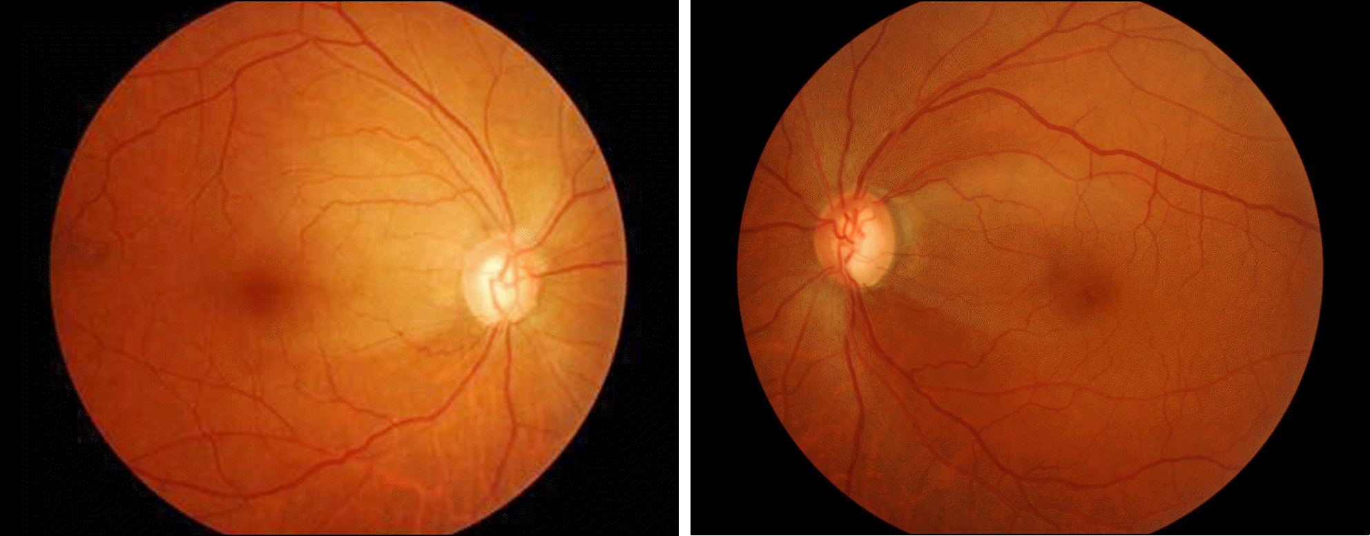
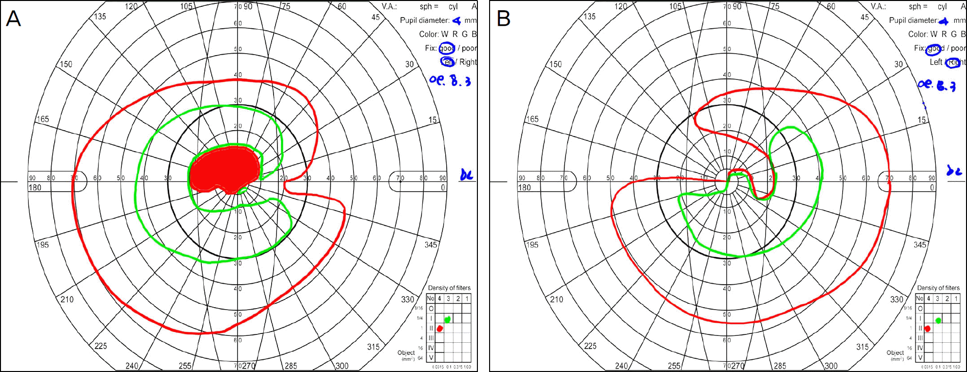
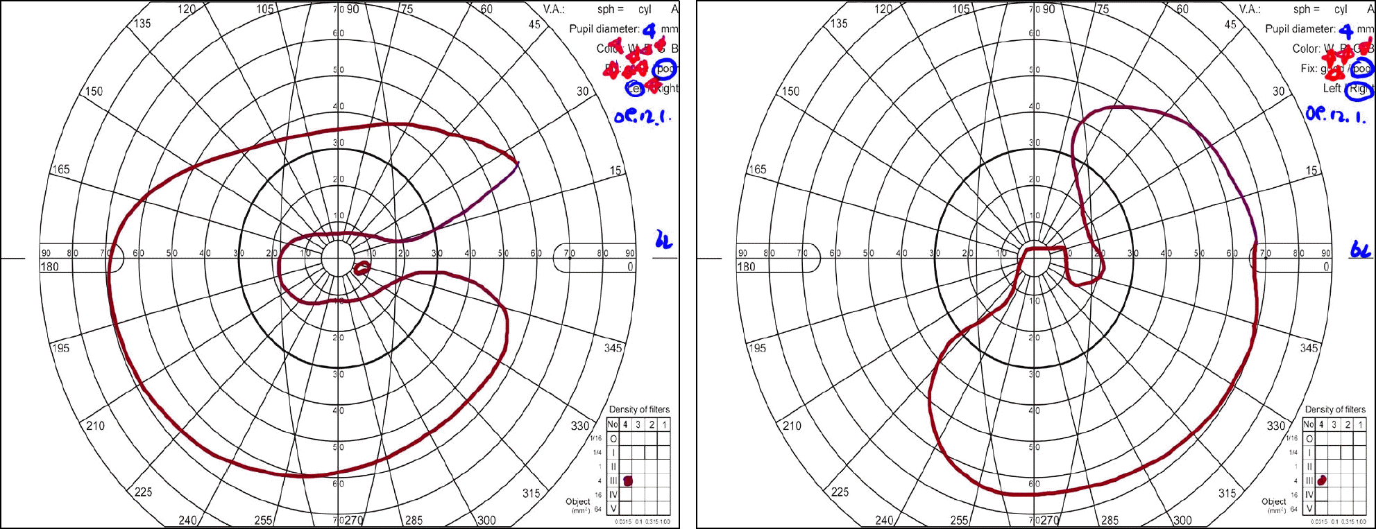
A 48-year-old man presented with complaints of decreased visual acuity in his left eye. His corrected visual acuity was 20/16 in the right eye and 20/63 in the left eye. The fundus photographs revealed a bilateral, superotemporal and inferotemporal retinal nerve fiber layer defect, corresponding to his visual field defect. The patient was diagnosed with bilateral NTG. After 2 months, the patient's corrected visual acuity in the left eye worsened to counting fingers and a central visual field defect was noticed in the Humphrey visual field test in the left eye. At 4 months after the initial visit, his corrected visual acuity in the right eye became 20/100, and the Goldmann visual field test demonstrated cecocentral scotoma. The fundus photographs showed a papillomacular bundle defect in his left eye. At 7 months after the initial visit, his visual acuity was hand movement in the right eye and a finger count in the left eye. A series of LHON gene mutation tests revealed a 11778 mitochondrial gene mutation, and the patient was diagnosed with LHON.
References
1. Leber T. Ueber hereditare und congenital angelegte Sehnervenleiden. Alberecht Von Graefes Arch Klin Exp Ophtalmol. 1871; 17:248–91.
2. Wallace DC, Singh G, Lott MT. Mitochondrial DNA mutation associated with Leber's hereditary optic neuropathy. Science. 1988; 242:1427–30.

3. Hwang JM, Jung YC. The etiology of optic neuropathy. J Korean Ophthalmol Soc. 1999; 40:1078–83.
4. Hwang JM, Park HW. A mitochondrial mutation in Leber's hereditary optic neuropathy. J Korean Ophthalmol Soc. 1995; 36:153–8.
5. Quigley HA, Addicks EM, Green WR. Optic nerve damage in human glaucoma. III. Quantitative correlation of nerve fiber loss and visual field defect in glaucoma, ischemic neuropathy, papilledema, and toxic neuropathy. Arch Ophthalmol. 1982; 100:135–46.
6. Von Graefe A. Über die iridectomie bei glaucom und über den glaucoma usen prozess. Albrecht von Graefes Arch Ophthalmol. 1857; 3:456–650.
7. Choe YJ, Hong YJ. The prevalence of glaucoma in Korean careermen. J Korean Ophthalmol Soc. 1993; 34:153–8.
8. Kwak HW, Joo MJ, Yoo JH. The significance of fundus photography without mydriasis during health mass screening. J Korean Ophthalmol Soc. 1997; 38:1585–9.
9. Newman NJ, Lott MT, Wallace DC. The clinical characteristics of pedigrees of Leber's hereditary optic neuropathy with the 11778 mutation. Am J Ophthalmol. 1991; 111:750–62.

10. Lauer SA, Ackerman J, Sunness J, et al. Leber's optic atrophy with myopia masquerading as glaucoma: case report. Ann Ophthalmol. 1985; 17:146–8.
11. Opial D, Boehnke M, Tadesse S, et al. Leber's hereditary optic neuropathy mitochondrial DNA mutations in normal-tension glaucoma. Graefes Arch Clin Exp Ophthalmol. 2001; 239:437–40.

12. Mashima Y, Kimura I, Yamamoto Y, et al. Optic disc excavation in the atrophic stage of Leber's hereditary optic neuropathy: comparison with normal tension glaucoma. Graefes Arch Clin Exp Ophthalmol. 2003; 241:75–80.

13. Nikoskelainen E, Hoyt WF, Nummelin K. Ophthalmoscopic findings in Leber's hereditary optic neuropathy. II. The fundus findings in the affected family members. Arch Ophthalmol. 1983; 101:1059–68.
14. Hwang JM, Chang BL, Park SS. Clinical Manifestations of Leber's Hereditary Optic Neuropathy with 11778 mitochondrial DNA Mutation in Koreans. J Korean Ophthalmol Soc. 2000; 41:1775–81.
Figure 1.
Retinal nerve fiber layer and disc photographs of the patient at the initial visit. Note superotemporal and inferotemporal retinal nerve fiber layer defect and disc rim notching in both eyes. Disc hyperemia or pseudoedema is not observed.
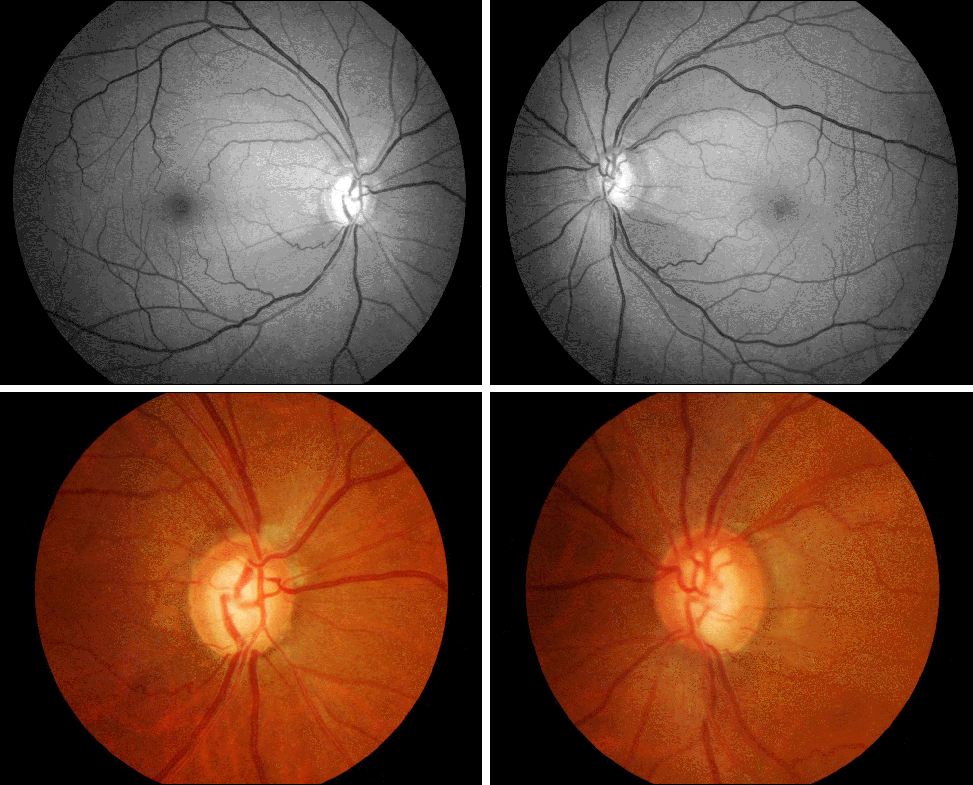
Figure 2.
Humphrey visual field (HVF) test. (A) Note central visual field defect in the left eye. (B) Arcuate visual field defect is observed in the right eye.
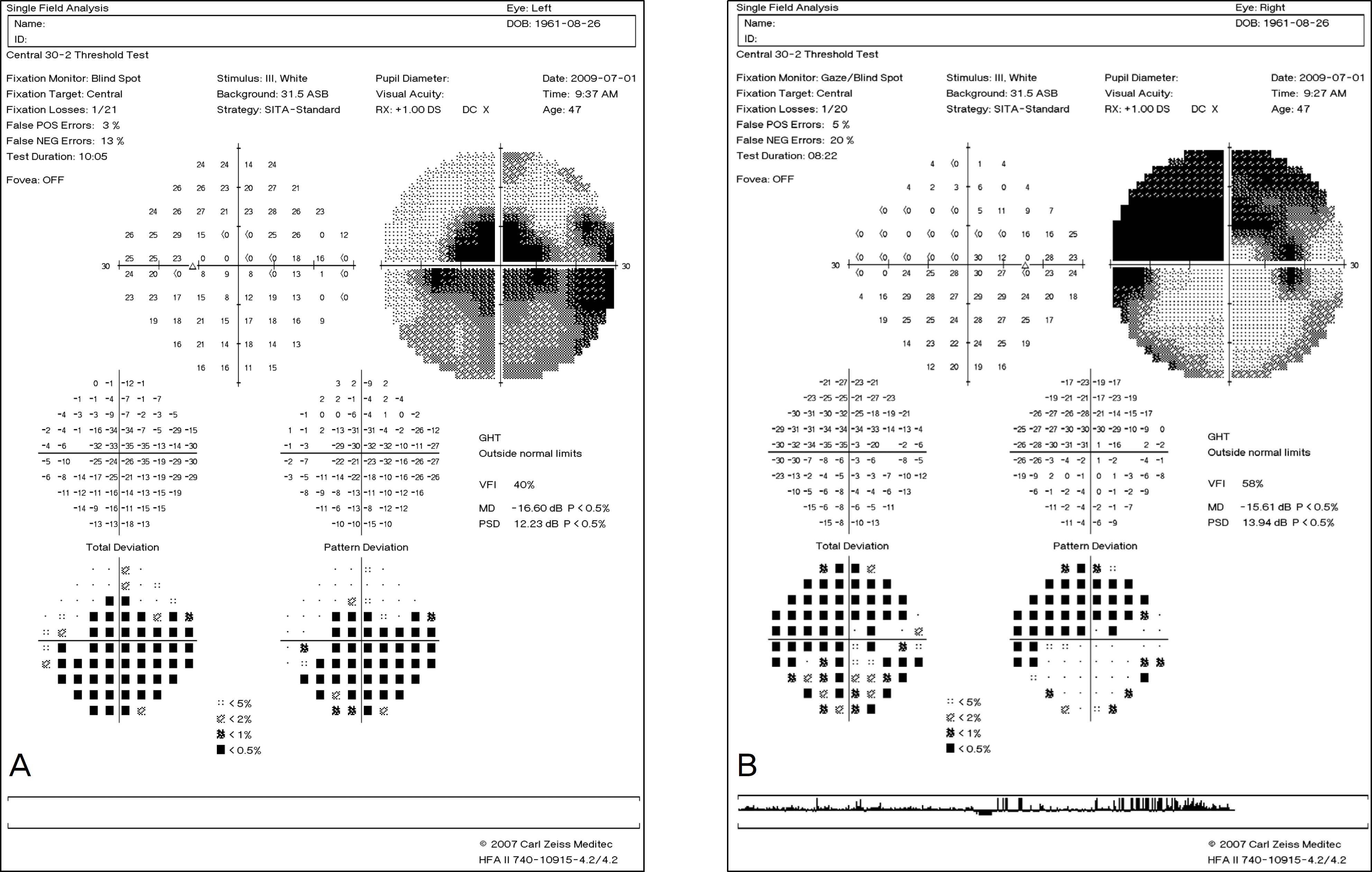
Figure 3.
Fundus photographs of the patient 4 months after the initial visit. Note a newly appeared papillomacular bundle defect in the left eye.


 PDF
PDF ePub
ePub Citation
 Print
Print


 XML Download