

 PDF
PDF ePub
ePub Citation
 Print
Print


INTRODUCTION
The laboratory identification of pathogenic yeasts is very important because of the increasing incidence of opportunistic infections and the rise in resistant strains of yeasts. The widespread use of immunosuppressive agents or broad-spectrum antibiotics predisposes patients to subsequent opportunistic fungal infections. The majority of these infections are caused by Candida albicans, but in recent years, close to 50% of all episodes of candidemia have been caused by non-albicans species [1-3]. The incidence of candidemia due to these uncommon Candida spp. appears to be increasing, and certain species, such as C. dubliniensis, have been reported to be less susceptible or resistant to antifungal agents [4].
C. dubliniensis was first isolated in 1995 from the oral cavities of HIV-infected individuals and AIDS patients [5]. While the clinical significance of C. dubliniensis seems to involve its association with HIV infection, C. dubliniensis has also been increasingly found in individuals who are not infected with HIV [6, 7]. Identification of C. dubliniensis can be problematic in routine clinical practice due to its phenotypic resemblance to C. albicans [8]. Because there have been no prior case reports of C. dubliniensis candidemia in Korea, the prevalence of this species among the Korean population remains unclear. In this article, we report the first case of C. dubliniensis candidemia in Korea, which was identified using both phenotypic and genotypic methods.
CASE REPORT
A 64-yr-old woman who presented with partial seizures, drowsiness, and recurrent fever was transferred to our hospital with a central venous catheter and L-tube. She had been treated for fever with ceftriaxone at another hospital, but had shown no clinical improvement. On admission to our hospital, the patient, who also had a medical history of hypertension, had a body temperature higher than 39℃. Viral meningitis and bacterial pneumonia were immediately suspected. Laboratory investigation revealed a leukocyte count of 5,920/mm3 with 87% neutrophils, 10 g/dL of hemoglobin, and a platelet count of 136,000/mm3. The serum level of C-reactive protein was elevated (270.72 mg/L). The patient underwent cerebrospinal fluid (CSF) tap, chest X-ray examination, and two sets of peripheral blood cultures, and was started on an empirical antibiotic combination of ticarcillin/clavulanate for the bacterial pneumonia and on acyclovir for the suspected viral meningitis for 4 days. However, this treatment yielded no clinical improvement. More than 3 days after admission, the patient's fever persisted, and her mental status change was exacerbated by convulsions. The patient continued to have fevers of >39℃ and was subsequently transferred to a medical intensive care unit (ICU).
The blood cultures obtained to evaluate the fever on the first day of admission were negative. Three days after admission, a second blood culture was performed and microbial growth was observed after 24 hr of incubation in the BacT/Alert 3D system (bioMerieux, Durham, NC, USA). The same microorganisms were found in culture sets of central venous catheter tips, in bile cultures, and in a third blood culture taken 3 days after the second blood culture. A subculture on sheep blood agar plates was performed for the peripheral blood and other specimens. After 24 hr of incubation on sheep blood agar plates at 35℃ in the presence of 5% CO2, we observed smooth and white colonies, suspected to be Candida spp. Isolates were subcultured on Sabouraud dextrose agar (SDA) plates for further phenotype testing. C. albicans ATCC 14053 and C. dubliniensis ATCC MYA-646 were used as reference strains.
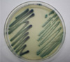
Identification was made on the basis of a combination of various phenotypic methods and confirmed by species-specific multiplex PCR. Isolates produced germ tubes in human pooled serum in 3 hr at 37℃ (Fig. 1) and did not grow at 45℃ for 96 hr on SDA plates. Isolates were cultured on CHROMagar Candida media (Becton Dickinson, Sparks, MD, USA) at 35℃ for 48 hr. The resulting colonies were dark green in color, which differs from the light to medium green color of the more commonly observed C. albicans (Fig. 2). These isolates were identified as C. dubliniensis using the Vitek 2 ID-YST System (bioMerieux Inc., Hazelwood, MO, USA) (probability, 91.0%) and the latex agglutination test using Bichro-Dubli Fumouze (Fumouze Diagnostics, France) (Fig. 3) [9]. To confirm this finding, we performed species-specific multiplex PCR using the Seeplex Candida Detection kit (Seegene, Seoul, Korea), which contained two sets of primer mixtures for the detection of 8 different Candida spp. [10]. The isolates were confirmed as C. dubliniensis.
An antifungal susceptibility test was performed using the Vitek 2 system (bioMerieux Inc.), and we determined that the isolates were susceptible to flucytosine, fluconazole, voriconazole, and amphotericin B. On the basis of the results of the susceptibility tests, the patient was treated with fluconazole for 4 weeks. In addition, the central venous catheter that was the suspected cause of the candidemia was removed immediately after C. dubliniensis was identified from the second blood culture. After initiation of antifungal therapy and removal of the central venous catheter, the patient's clinical symptoms, including the fever, were gradually improved. However, due to other medical problems including cholecystitis, gastric ulcer, and hypertension, the patient remained in the ICU for 93 days, after which she was discharged to another hospital.
DISCUSSION
The incidence of candidemia in the overall population ranges from 1.7 to 10 episodes per 100,000 inhabitants, and Candida is one of the 10 leading causes of bloodstream infections in developed countries. An estimated 33-55% of all episodes of candidemia occur in the ICU environment and are associated with mortality rates ranging from 5% to 71% [3]. Candidemia may have an endogenous or an exogenous origin, and in recent years, a growing proportion of episodes of candidemia have been caused by Candida spp. other than C. albicans. There are a number of conditions predisposing ICU patients to candidemia, including prior abdominal surgery, intravascular catheters, acute renal failure, malignancy, vancomycin treatment longer than 3 days, parenteral nutrition, broad-spectrum antibiotics, a prolonged ICU stay, the use of corticosteroids, and mucosal colonization with Candida spp. [3, 11, 12]. For the patient in this case, the predisposing factors included a central venous catheter, parenteral nutrition, broad-spectrum antibiotics, and a prolonged ICU stay. Among these, the central venous catheter was the most likely explanation for nosocomial candidemia, which is supported by the clinical improvement of the patient after removal of the catheter. A central venous catheter has also been cited as a risk factor for nosocomial candidemia in several reports, and removal of catheters has been recommended to reduce the rate of catheter-related bloodstream infection and to prevent nosocomial clusters in the ICU [13, 14].
C. dubliniensis has recently been described as an opportunistic yeast that is primarily associated with oropharyngeal infections in HIV-infected patients. Unlike C. albicans, C. dubliniensis is rarely found in the oral microflora of normal healthy individuals, and it is responsible for as few as 2% of cases of candidemia (compared to approximately 65% for C. albicans) [15].
Phenotypically, C. dubliniensis and C. albicans have many similarities, including their microscopic morphology and ability to form germ tubes in serum [8]. Therefore, the objective of most previous studies has been to distinguish C. dubliniensis from C. albicans or other germ tube-positive Candida species [16, 17]. Tests based on different phenotypic characteristics, such as the inability of C. dubliniensis to grow at 45℃ [18], and/or in Sabouraud dextrose broth containing 6.5% sodium chloride (NaCl) [19]; the production of abundant pseudohyphae and chlamydospores on Staib agar [20]; and the formation of dark green colonies on CHROMagar Candida medium [21], have been claimed to differentiate between the two species. However, these characteristics are not definitive, and identification of C. dubliniensis in routine laboratory testing remains a problem. Additional tests, such as the latex agglutination test and molecular assays, are helpful in accurately differentiating between the two species.
The latex agglutination test used for this case is a commercialized protocol for identifying C. dubliniensis isolates that uses particles coated with a monoclonal antibody, which allows for the specific detection of an antigen located on the surface of the C. dubliniensis blastoconidia [9]. The latex agglutination of C. dubliniensis is visible to the unaided eye. Molecular techniques employing PCR have also increased the accuracy of identifying uncommon Candida spp. Although 16S RNA gene sequencing is a useful and definitive method for most Candida spp., it requires a specialized instrument to analyze the product. Multiplex PCR using a specific primer can serve as an alternative, easy to perform, and accurate diagnostic tool for the identification of C. dubliniensis [10, 22, 23].
Resistance to antifungal agents is rarely found in Candida spp. The fluconazole resistance rate was reported to be 2% in Korean patients with candidemia, and each of the Candida spp. that caused candidemia in that study showed susceptibility to voriconazole [24, 25]. However, uncommon Candida spp., such as C. guilliermondii, C. rugosa, and C. dubliniensis, had susceptible-dose-dependent minimum inhibitory concentration (MIC) values for fluconazole [4]. Resistance to fluconazole has been reported to occur only rarely in C. albicans (0.6%), while occurring more commonly in C. dubliniensis (3.1%) [2]. However, the vast majority of C. dubliniensis isolates identified to date are susceptible to all of the commonly used antifungal agents [13]. C. dubliniensis isolates in the present case showed susceptibility to flucytosine, fluconazole, voriconazole, and amphotericin B, which corresponds with previous reports.
In this report, we describe the case of C. dubliniensis candidemia in an ICU patient with pneumonia and meningitis. To our knowledge, this report is the first case of C. dubliniensis candidemia in Korea and the first case identified phenotypically and subsequently confirmed by molecular methods. During the diagnostic process, a C. dubliniensis infection should be distinguished from infection caused by other Candida spp., particularly C. albicans. Confirmation through various methods, such as latex agglutination and multiplex PCR, is needed for the accurate identification of isolates. Both clinicians and microbiologists should be aware of predisposing factors for C. dubliniensis candidemia to promote early diagnosis and appropriate treatment.


 XML Download