

 PDF
PDF ePub
ePub Citation
 Print
Print


INTRODUCTION
Pilar sheath acanthoma is a rare benign follicular hamartoma. Mehregan and Brownstein (1) first reported 9 cases in 1978 that included a spectrum of lesions composed of lobules of follicular epithelium with infundibular maturation. To date, approximately 50 cases have been described worldwide, and only 5 cases have been reported in Korean dermatology journals until 2009 (2). In 1993, Ackerman et al. (3) investigated 21 cases and discovered that in addition to infundibular differentiation, isthmus, and, rarely, sebaceous ducts, apocrine glands, the inner root sheath, and bulb differentiation may be involved. Therefore, Hurt suggested that “pilar sheath acanthoma” should be renamed “lobular infundibuloisthmicoma”. To the best of our knowledge, despite many reports on the clinicopathological aspects of pilar sheath acanthoma, this entity has not been well described in the radiologic literature, and ultrasound (US) findings have not been documented. Here, we report the first US findings of pilar sheath acanthoma located on the medial aspect of the calf.
CASE REPORT
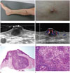
A 56-year-old male patient was hospitalized for treatment of a painful palpable nodule with a central pore-like opening on the medial aspect of the right calf. He had undergone surgery for treatment of varicose veins on the right leg 3 years ago, and then he experienced pain radiating from the operation site to the right toes. Since the past 10 months, a solitary erythematous hyperpigmented papule measuring approximately 1 cm with pus discharge was visible on the medial aspect of the right calf (Fig. 1A, B). On gross examination, the clinical differential diagnosis included an epidermoid cyst, an open comedo, or a dilated pore of Winer.
US was performed to identify the location and to characterize the nodule. A well-defined oval hypoechoic nodule, which measured about 0.65 × 0.35 cm, with hypoechoic capping within the dermis over the medial aspect of the right calf was noted on gray scale US (Fig. 1C). This hypoechoic nodule communicated with hypoechoic capping by a narrow hypoechoic neck. The gray scale US also revealed no posterior acoustic enhancement. This lesion showed internal hypervascularity on color Doppler images (Fig. 1D). According to US findings, the provisional diagnosis was an epidermoid cyst or pilomatricoma.
An excisional biopsy was performed to confirm the diagnosis. Histopathological examination revealed a lobular patulous tumor that was connected to the epithelium, and tumor cells consisting of mixed blue-gray (infundibular) and pink (isthmic) corneocytes were noted in the dermis. The findings were compatible with pilar sheath acanthoma (Fig. 1E, F).
DISCUSSION
Pilar sheath acanthoma is a rare benign follicular hamartoma. Middle-aged and elderly individuals are commonly affected. Pilar sheath acanthoma is characterized by a small (5 to 10 mm in diameter), solitary, painless, skin-colored papule located on the head or neck, particularly around the upper lip (4). A central pore that is occasionally plugged with keratin is often present (4).
Diseases that should be considered in the clinical differential diagnosis include an epidermoid cyst, an open comedo, trichofolliculoma and a dilated pore of Winer. From the pathological viewpoint, it is difficult to differentiate pilar sheath acanthoma from trichofolliculoma and a dilated pore of Winer due to similar histopathological findings. A common histopathological feature among these tumors is the central sinus, which contains keratinous material, and is lined by epithelium that is continuous with the surface epidermis (5). In pilar sheath acanthoma, abortive hair follicle-like structures are present but do not show a high degree of differentiation; hair shafts are absent within the central cavity, prominent fibrovascular stroma is lacking, and several small cysts within the mass of the cyst wall are visible. In trichofolliculoma, small hair follicles radiate from the wall of the central infundibular cyst (6). A dilated pore of Winer is a patulous follicle that contains hair. Numerous small digitate projections radiate from the follicular epithelium into the surrounding connective tissue (45). Mehregan suggested that pilar sheath acanthoma is less mature than a dilated pore of Winer but more mature than a tumor of the follicular infundibulum (1). Treatment consists of surgical excision or electrodesiccation and curettage (4).
In this case, the US findings revealed a non-specific soft tissue nodule that was a well-defined, hypoechoic nodule with hypoechoic capping and had hypervascularity within the dermis. According to the radiologic-pathologic correlations, a lobular patulous tumor that was connected to the epithelium was noted in the dermis, and it had a central pore-like opening that exhibited a hypoechoic nodule with a narrow hypoechoic neck and erosive or ulcerative epidermis with hypoechoic capping on US. When there are superficial soft-tissue masses, the possible diagnosis is variable. Superficial soft-tissue masses may be classified into one of the following general diagnostic categories: mesenchymal tumors, skin appendage lesions, metastatic tumors, other tumors and tumorlike lesions, and inflammatory lesions (7). Also, it is important to identify the exact location of the mass in the differential diagnosis (7). Skin appendage lesions, which include an epidermoid cyst, pilomatricoma, cystadenoma, cylindroma, and syringoma, originate in the epidermis and dermis (7). In this case, US revealed a well-defined oval hypoechoic nodule with hypoechoic capping within the dermis. Thus, we considered that the possible diagnosis was an epidermoid cyst or pilomatricoma. The epidermoid cyst is filled with loosely packed lamellae of keratin; the walls resemble those of a follicular infundibulum (8). At US, the cyst appears as a circumscribed circular or oval hypoechoic mass, often in association with a hair follicle (7). Lee et al. (8) reported the US findings of an epidermoid cyst, and they stated that 96% of the lesions showed posterior sound enhancement and 83% of the lesions showed no signal on color Doppler image. Pilomatricoma is a benign superficial tumor of the hair follicle (9). It is the most common solid cutaneous tumor in patients aged 20 years and younger. US features of pilomatricoma are a well-defined oval mass with strong posterior acoustic shadowing at the junction of the dermis and subcutaneous fat with focal thinning of the overlying dermis (10). Hwang et al. (9) reported that US revealed a well-defined oval hyperechoic nodule with posterior acoustic shadowing. The reason for posterior acoustic shadowing is probably that pilomatricoma shows frequent calcification. Thus, we can differentiate pilar sheath acanthoma from epidermoid cyst or pilomatricoma by some US features, such as posterior sound enhancement or posterior acoustic shadowing.
Pilar sheath acanthoma can be considered a possible diagnosis when a well-defined dermal nodule with a central pore-like opening is a hypoechoic nodule with hypoechoic capping and it shows hypervascularity on US images.
 XML Download