

 PDF
PDF ePub
ePub Citation
 Print
Print


INTRODUCTION
In addition to function, smile esthetics is considered as high priority during the planning stage of orthodontic treatment, with the goal of delivering a healthy, natural, and confident smile. One of the most important goals of smile design is to achieve posterior tooth display, allowing filling of the buccal corridors.12345 In order to reach this goal, it is necessary that the patient exhibits an optimal transverse dimension of maxillary dentoalveolar bone, as well as appropriate buccolingual inclinations of the posterior teeth.67891011 This is necessary for both functional and esthetic occlusion. Thus, the torque values of the posterior brackets are important in achieving this goal and must interface favorably with the lateral and protrusive forces.
Studies that have investigated inclinations of posterior teeth have often grouped subjects according to sagittal or vertical skeletal characteristics. Shu et al.12 compared groups assigned according to sagittal characteristics and found that Class II division 1 subjects showed more lingually inclined maxillary molars, compared with individuals with Class I occlusion. In contrast, they could not find any difference for mandibular molars. Shu et al.12 thus suggested that the transverse disharmony of the arches in Class II division 1 cases results from inclination of the maxillary teeth; however, there was no vertical classification or discussion of the effect of vertical characteristics. Ahn et al.13 found more lingual inclination in the mandible and more buccal inclination in the maxilla in Class III subjects, when compared with Class I subjects. Their results showed that this finding was correlated with ANB (A point, nasion, B point) angle. Importantly, there was no mentioning of the vertical characteristics of the subjects in this study, either.
Studies have shown varying results regarding the buccolingual inclination of the posterior teeth in relation to vertical growth type.141516171819 Tsunori et al.14 measured the mandibular buccolingual inclinations of hyper- and hypodivergent groups comprising Class I or Class II cases; they concluded that the hyperdivergent group exhibited more buccally inclined posterior teeth than the hypodivergent group. In contrast, Janson et al.15 found that maxillary molars of hyperdivergent Class I and Class II division 1 subjects were buccally inclined, relative to those of hypodivergent Class II division 2 subjects; however, there was no such difference in a comparison of mandibular molars. Ross et al.16 found no statistically significant difference in molar inclinations among different vertical facial types. Masumoto et al.17 evaluated mandibular molars in a series of Japanese dry skulls showing normal occlusion and found that in the hypodivergent group, second molars had more lingual inclination than in the hyperdivergent group. Grosso et al.,18 who only classified the subjects according to vertical facial type, demonstrated that the maxillary and mandibular molars of subjects in the hyperdivergent group were lingually inclined.
The heterogeneity of results in the literature, mainly related to classifying patients according to either sagittal or vertical characteristics, creates a challenge for the clinician in determining the characteristic inclinations of the posterior teeth in a specific patient; therefore, the purpose of this study was to evaluate the buccolingual molar inclinations of maxillary and mandibular arches, specifically in skeletal Class I patients with different vertical facial type.
MATERIALS AND METHODS
The sample for this study was generated by retrospective screening of three-dimensional cone-beam computed tomography (CBCT) images in the archives of the Oral Radiology Department of Yeditepe University Dental School, acquired between January 2008 and January 2014. The inclusion criteria were as follows: subjects aged 20 to 45 years, who exhibited a Class I maxillomandibular relationship, no facial asymmetries, no cleft lip or palate, no impacted or missing teeth in the measurement site, no periodontal disease, no diagnosed systemic diseases, and no craniofacial dysmorphology. Patient data were handled according to the requirements and recommendations of the Declaration of Helsinki. Ethical approval (no. 207) was obtained from the institutional review board of Yeditepe University. The images used in this study were acquired at 120 kVp and 3.8 mA, with an exposure time of 40 seconds; they were created with a focal spot of 3.3 mm and a voxel size of 0.093 mm on a CBCT unit (Iluma; IMTEC Corporation, Ardmore, OK, USA). The images were saved as Iluma vision viewer files.
Cephalometric analyses were performed on CBCT data to reveal sagittal and vertical skeletal characteristics of the subjects. Class I subjects with an ANB angle of 0° to 4° were included in the study. Sella-nasion/gonion-menton (S-N/Go-Me) angle and S-Go/N-Me ratio were used to assign the subjects into groups according to vertical growth patterns. S-N/Go-Me angle of < 27° indicated hypodivergency, 27° to 37° indicated normodivergency, and > 37° indicated hyperdivergency.20 For S-Go/N-Me; a ratio of < 61% indicated hyperdivergency, 61% to 69% indicated normodivergency, and > 69% indicated hypodivergency.21 Subjects who did not fulfill these criteria were excluded from the study. Ultimately, CBCT records of 135 patients were included in the study. The distribution of the patients into groups is provided in Table 1. The normodivergent group consisted of 46 subjects (24 females, 22 males) with a mean age of 30.2 ± 6.3 years; the hypodivergent group consisted of 49 subjects (26 females, 23 males) with a mean age of 30.3 ± 7.6 years; and the hyperdivergent group consisted of 40 subjects (24 females, 16 males) with a mean age of 29.5 ± 5.3 years.
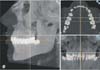
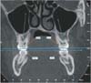
The images were reoriented in three planes of space. The anatomical occlusal plane was aligned parallel to the floor in the sagittal view. In the coronal and axial views, CBCT images were adjusted using a line passing through the buccal cusps of the maxillary first molars (Figure 1). A maxillary occlusal plane was then constructed between the central sulci of the maxillary right and left first molars on the coronal slice showing the bifurcation of both molars. Maxillary first and second molar buccolingual inclinations were measured as the inner angles formed by the long axes (a line passing by the central sulcus and bifurcation) of the teeth, relative to the maxillary occlusal plane (Figure 2). For the mandibular teeth, the mandibular occlusal plane was constructed between the central sulci of the mandibular right and left first molars on the coronal slice showing the apices of both molars. Mandibular first and second molar buccolingual inclinations were measured as the inner angles formed by the long axes (a line passing by the central sulcus and apex) of the teeth, relative to the mandibular occlusal plane (Figure 2).
Statistical analyses were performed with NCSS 2007 statistical software (NCSS, Kaysville, UT, USA). Descriptive statistics, including the means and standard deviations, were obtained for the data. The normal distribution of the data was assessed using the Shapiro–Wilk test. All variables were normally distributed. One-way analysis of variance was used for intergroup comparisons. Independent t-tests were used to investigate sex differences. The results were evaluated at the p < 0.05 significance level, with a 95% confidence interval.
One week after the first measurements, buccolingual molar inclination measurements were repeated by the same author (M.T.). Systemic error was calculated using intraexaminer reliability values, which were determined via intraclass correlation coefficients.
RESULTS
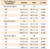
Intraclass coefficients were between 0.883 and 0.997, indicating that the operator was consistent during repeated measurements. There was no statistically significant difference between females and males in terms of buccolingual molar inclinations (Table 2). Therefore, the data were pooled. The mean buccolingual inclination of the maxillary first molars of the hyperdivergent group was 1.6° larger than that of the normodivergent and hypodivergent groups, with a possible trend toward significance. However, no statistically significant difference was found in maxillary and mandibular first and second molar inclinations of the normodivergent, hypodivergent, and hyperdivergent subjects (p = 0.057, 0.370, 0.148, and 0.081, respectively) (Table 3).
DISCUSSION
The determination of the smile components in a patient is very important in designing a successful orthodontic treatment plan.22 Posterior components of the smile include the inclinations of the posterior teeth, which have an important effect on esthetics and proper function. The current literature consists of studies where vertical and sagittal characteristics of the compared groups are disorganized, creating confusion and resulting in heterogeneity. In a study where the buccolingual inclinations of posterior teeth with different facial patterns were evaluated, the hyperdivergent group comprised of both skeletal Class I and Class II division 1 cases, whereas the hypodivergent group consisted of Class II division 2 cases.15 Notably, molar inclinations may be affected by the sagittal skeletal discrepancy.1213 There have been studies involving the sole description of sagittal characteristics, with no comment on vertical relationships; however, the effect of vertical growth on molar inclinations may have considerable relevance.121322 The present study, therefore, describes the molar inclinations in Class I patients presenting either hypo-, hyper-, or normodivergent vertical patterns.
Analysis of the inclination of maxillary molars in hypo-, hyper-, or normodivergent Class I patients showed that there were no differences among the groups. As in our study, Ross et al.16 and Grosso et al.18 also found no statistically significant differences among various facial types. Importantly, Ross et al.16 compared the molar inclinations in hyperdivergent, normodivergent, and hypodivergent subjects using study models for measurements, irrespective of skeletal sagittal pattern. Grosso et al.18 used a different approach, where they measured both long axis and buccal surface inclinations of the maxillary first molars, relative to the occlusal plane, on CBCT images in three vertical groups, thus revealing similar angulations among different facial types.
Conversely, Janson et al.15 found that the hyperdivergent group showed higher buccal maxillary molar inclination values than the hypodivergent group. In their hyperdivergent group, there were both Class I and Class II division 1 patients. Furthermore, their low angle group consisted of Class II division 2 subjects. Since there is evidence for a difference in the inclination of the maxillary molars between Class I and Class II subjects,12 the results of the study by Janson et al.15 are questionable.
In our study, the mean mandibular first molar inclinations were not statistically significantly different. Even though the numerical inclination values of the mandibular molars found in the studies by Janson et al.15 and Ross et al.16 were not comparable with our numerical data, these prior studies showed no difference of inclination values for mandibular molars in different vertical facial types, supporting our findings. However, Grosso et al.18 measured inclination not only from the long axis of the teeth, but also from a line drawn to the buccal surface of the clinical crown. Their study revealed an increase of lingual inclination in dolichofacial subjects as measured from the crowns, but not from the axes of the teeth; it also showed a buccolingual height difference in the mandibular first molars among the groups. Similarly, Masumoto et al.,17 while not detecting any difference in the inclination of the first molars, found that the lower second molars of the hyperdivergent group were more lingually inclined. However, there was no sagittal description of the dry skulls of Japanese ethnicity that demonstrated normal occlusion with minimal discrepancy, without crossbite or facial asymmetry. In another dry skull study by Tsunori et al.,14 the hypodivergent group had more lingually inclined molars, contrary to the previous study. This difference may be due to the inclusion and pooling of both Class I and Class II sagittal patterns, and may be related to the weak musculature of the hyperdivergent patients. However, we suspect that the lack of description of sagittal and transversal relationships may have played an important role in the results.
The limitations of this study include the possibility of bias in the execution of the study and handling of the data, as well as the sole evaluation of Class I subjects. Further studies performed on adults, considering both sagittal and vertical characteristics separately, may provide a healthier and more meaningful discussion regarding what can be done for normo-, hyper-, and hypodivergent Class I, Class II, and Class III subjects.
CONCLUSION
There was no statistically significant difference between females and males in terms of buccolingual molar inclinations. Furthermore, there was no statistically significant difference in the molar inclinations of hyperdivergent, normodivergent, and hypodivergent adult subjects with Class I sagittal relationships. However, maxillary first molars of the hyperdivergent subjects appeared to be approximately 2° more upright than those of the other subjects.


 XML Download