

 PDF
PDF ePub
ePub Citation
 Print
Print


Introduction
Marbofloxacin is a fluoroquinolone antibiotic developed only for application in veterinary medicine [14] that acts by inhibiting bacterial DNA-gyrase. It has been demonstrated that marbofloxacin is potent in vitro against Mycoplasma, most Gram-negative and some Gram-positive bacteria, and some intracellular pathogens, but has limited or little activity against anaerobes [316]. Most porcine respiratory pathogens, such as Actinobacillus pleuropneumoniae and Haemophilus parasuis, are very sensitive to marbofloxacin. Currently, in China, marbofloxacin has been not licensed for use in pigs. However, because of its spectrum of activity against swine pathogens, it is being used in an extra-label manner to treat pigs' respiratory diseases and sows' metritis0–mastitis0–agalactia syndrome.
Similar to other fluoroquinolones, marbofloxacin has a low plasma protein binding rate [7], a large volume of distribution with potent activity [69], and high drug concentrations in tissues and body fluids [1]. The pharmacokinetics of marbofloxacin in plasma and urine has been studied in pigs under different administration routes [14], and it was shown that marbofloxacin is well absorbed with a bioavailability of 91.5% after intramuscular administration, and its body clearance rate decreases significantly with pig age after intravenous administration. Another study showed easy penetration of marbofloxacin into pig tonsils [19]. In addition, a pharmacokinetics study in pigs' tissue cage fluid demonstrated efficient distribution of marbofloxacin into tissues [5]. Some studies have been published regarding the tissue residues of marbofloxacin in other species [101221]. However, there is a paucity of systematic information in the literature regarding the distribution of marbofloxacin in the respiratory system. Moreover, there is also a paucity of literature regarding the distribution of marbofloxacin in the edible tissues of pigs.
Information about tissue distribution of an antibiotic would be very valuable for improving treatment efficacy and avoiding problems associated with residues. Therefore, the purpose of this study was to investigate marbofloxacin's tissue distribution in pigs after a single intramuscular administration.
Materials and Methods
Animals
A total of 40 eight-week-old castrated cross-bred (Duroc × Landrace × Yorkshire) pigs weighing 20 to 23 kg were used in the study. The pigs were allowed to acclimate for seven days with free access to water and a drug-free pelleted diet. Five days before the start of the study, a complete wellness examination including a physical examination and blood samples was performed. Blood was collected from each pig, and a complete blood count (CBC) and chemistry panel (CP) were performed. No clinically significant abnormalities were noted on examination and all blood parameters were within normal ranges. The study (animal study protocol 201506007) was approved by the Institutional Animal Care and Use Committee (IACUC) of Henan University of Science and Technology. All animals were humanely handled.
Chemicals and reagents
Commercial marbofloxacin suitable for intramuscular injection (5 mL: 5 g, Lot No. 091008) was kindly provided by Hebei Yuanzheng Pharmaceutical (China). Marbofloxacin standard (99.82%, Lot No. H050408) was provided by the National Institutes for Food and Drug Control (China). Formic acid and acetonitrile of high-performance liquid chromatography (HPLC) grades were both purchased from Merck (Germany). The other reagents used in this study were all of analytical grades. Deionized water was purified by using a Milli-Q system (Millipore, USA).
Drug administration and sampling
Pigs were randomly divided into eight groups with five pigs in each group. Group 1 was used as a control. Pigs in the other groups were weighed and intramuscularly injected on the right side of the neck with marbofloxacin at a dosage of 2.5 mg/kg body weight. To obtain tissue and plasma samples, five pigs which received the intramuscular injection were randomly killed at 2, 6, 10, 24, 48, 72, and 96 h, respectively, whereas those in control group were sacrificed at 96 h. Samples of plasma and tissues, including muscle, lung, heart, kidney, liver, and muscle at the injection site, were collected from each animal. Tissue samples were thoroughly minced and stored at −20℃ prior to use.
Marbofloxacin determination
The marbofloxacin concentrations in plasma and tissues were measured by using a previously described method [20]. Briefly, 6 mL of trichloromethane was used to extract the marbofloxacin from 0.6 mL of plasma. Three grams of homogeneous tissue samples were extracted separately with 3 parts of trichloromethane (5 mL each) and 0.1 M sodium phosphate buffer (pH 7.4; 3 mL each). After shaking for 5 min and centrifugation at 3,180 × g for 10 min, the organic phase was pooled for tissues and collected for plasma, then evaporated using a stream of nitrogen at 30℃ and reconstituted in 1 mL mobile phase for tissues and 0.6 mL mobile phase for plasma.
The processed samples (50 µL) was injected into a Hypersil BDS-C18 column (4.6 mm × 250 mm, 5 µm; Elite Analytical Instruments, China) which was kept at 30℃. The mobile phases for HPLC analysis comprised 12.5% water, 12.5% acetonitrile, and 75% buffer which consisted of 1% formic acid and 0.5% triethylamine. Elution flow rate was set as 1 mL/min. An ultraviolet detector set to a wavelength of 295 nm was used to determine marbofloxacin presence.
Tissue kinetic analysis
Plasma and tissues concentrations versus time profiles were calculated based on mean marbofloxacin concentrations and non-compartmental modeling by using Phoenix WinNonlin (ver. 6.1; Pharsight, USA). The area under the concentration-time curve (AUC0–∞) was calculated by using a trapezoidal method. The peak concentration (Cmax) and time to reach Cmax (Tmax) were directly read from the concentration versus time profiles. The elimination rate constant (λz) was estimated by linear regression of mean drug concentrations versus time. The Cmax values in different tissues were compared by using SPSS (ver. 20.0; IBM, USA), and a multiple-range test was used to determine the significance of differences between mean concentrations. A p value lower than 0.05 was considered significant.
Results
The HPLC method was selective for marbofloxacin, and no endogenous interferences were found on chromatograms. The limit of quantitation (LOQ) for marbofloxacin was determined based on a signal-to-noise ratio greater than 10, and the LOQ values were 0.05 µg/g in tissues and 0.02 µg/mL in plasma. Marbofloxacin concentration was linear in tissues within the range of 0.050–5.00 µg/g (r > 0.998) and in plasma within the range of 0.020–5.00 µg/mL (r > 0.997). Mean recovery of marbofloxacin ranged from 90.82%0–96.43% in plasma and 84.36%0–90.27% in tissues. Repeatability was measured as within-run and between-run coefficients of variation, values of which were less than 7.52% in both plasma and tissues.
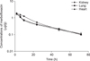
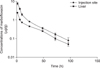
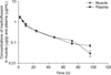
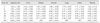
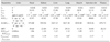
The concentrations of marbofloxacin in plasma and tissues are presented in Table 1 and Figs. 1, 2, 3. Marbofloxacin was detected at injection site, liver, muscle, and plasma up to 96 h after intramuscular administration and in kidney, lung, and heart up to 72 h after intramuscular administration. The highest concentration (7.64±1.06 µg/g) of marbofloxacin was measured in injection site, followed by liver (2.45±0.09 µg/g), kidney (1.93±0.06 µg/g), muscle (1.86±0.08 µg/g), lung (1.74±0.02 µg/g), heart (1.71±0.03 µg/g), and plasma (1.62±0.13 µg/mL). The kinetic parameters after a single intramuscular injection are listed in Table 2 for injection site, plasma, and each tissue type.
Discussion
Based on the plasma concentrations versus time data, a t1/2λz of 21.48 h was calculated for marbofloxacin when it was intramuscularly injected to pigs at 2.5 mg/kg body weight, a t1/2λz that is longer than those reported by Ding et al. [5] (17.3±5.38 h) and Schneider et al. [14] (15.10–15.4 h). The total body clearance of marbofloxacin decreases in older pigs [14], which may be the reason for the longer t1/2λz reported here. Pigs used here were eight weeks old and younger than those used by Schneider et al. [14]. In addition to age, differences between preparations of marbofloxacin used in the studies may be another reason for the inconsistent t1/2λz results. In addition to that in pigs, marbofloxacin pharmacokinetics have been investigated in other species. After intramuscular injection, the t1/2λz was 1.96, 3.15, 3.65, 7.16, 7.72, and 17.50 h in ostriches [4], ducks [8], sheep [15], camels [11], rabbits [13], and calves [17], respectively. These results indicate that the elimination of marbofloxacin from pigs is slower than that in other species. Such differences are relatively common and often associated with the following factors: breed, gender, age, body weight, disease, and heritable traits. Pharmacokinetic differences may also result from different eating habits, water intake, and exercise, which may lead to individual differences in blood flow. In a previous report [6], marbofloxacin was given orally to experimentally infected chickens, and its concentrations in tissues (except brain) exceeded those in plasma, which is consistent with the present results. A tissue residue study of marbofloxacin conducted in healthy chickens also indicated that marbofloxacin easily penetrated into all tissues [2].
The ratios of Cmax in tissues to that in plasma were between 1.05 and 3.17, and the AUC0–∞ values in tissues were also higher than that in plasma (1.06 to 4.71 times higher), indicating that marbofloxacin was easily distributed to tissues. The ratios of AUC0–∞ and Cmax values in tissues and plasma of infected chickens [6] were both higher than those observed in the present study of pigs. In addition to species difference, the presence of an infection perhaps affected concentrations by increasing the permeability of tissues, leading to more extensive distribution. After one single intramuscular injection, the highest marbofloxacin concentration (7.64±1.06 µg/g) was observed at the injection site, and, thereafter, the concentration presented a sustained downward trend. According to the report by Vilalta et al. [18], the intramuscular absorption rate constant of marbofloxacin ranged from 5.06 h−1 to 5.85 h−1 in pigs. This is consistent with the present results, which also indicate fast absorption of marbofloxacin. Based on the high residual concentrations in the injection site, the withdrawal time of marbofloxacin after intramuscular injection may be longer than that after oral administration. Moreover, the depletion of marbofloxacin residue after multiple intramuscular dosages should be studied to estimate further its withdrawal time in pigs.
When using fluoroquinolones to treat an infection, the ratio between AUC and minimum inhibitory concentration (AUC/MIC) and that between Cmax and MIC (Cmax/MIC) are both associated with successful therapeutic resolution. The MICs of marbofloxacin were reported to be 0.030–0.06 µg/mL and 0.0150–0.03 µg/mL against Actinobacillus pleuropneumoniae and Haemophilus parasuis, respectively [18]. According to the kinetic parameters determined in the present study, and the MIC data reported by Vilalta et al. [18], an application of 2.5 mg/kg marbofloxacin via intramuscular administration every 8 h in pigs could provide sufficient plasma concentrations to inhibit Actinobacillus pleuropneumoniae and Haemophilus parasuis. Such a multiple dosing regimen is suggested and should be followed in clinical veterinary medicine.
In conclusion, this study demonstrates that marbofloxacin is rapidDly absorbed, widely distributed, and slowly eliminated from pigs after a single intramuscular dose. Based on the derived kinetic parameters and MICs, intramuscular injection of marbofloxacin at 2.5 mg/kg per 8 h interval might be highly efficacious against susceptible bacteria in pigs.
 XML Download