

 PDF
PDF ePub
ePub Citation
 Print
Print


Introduction
Because of the rapid growth of intensive livestock production in recent years, indoor animal husbandry facilities now pose a potential health risk to husbandry workers and animals owing to the generation of inhalable particulate emissions and the harmful compounds adhered to them [6]. These organic dusts include bacteria, viruses, and fungi, as well as microbial secondary metabolites. Organic dusts, which are generated from bedding, fecal materials, animal skin, or feedstuffs, float into indoor air during cleaning or through animal activities such as feeding or feathering [61222]. Exposure to organic dusts may cause alteration of immune responses in animals and farm workers exposed [1832]. These health risks are heightened when large amounts of microorganisms are present in the air, which increases the risk of spreading diseases from one livestock building to another or to neighboring communities. In a study conducted in Vietnam, the bacterium (Enterococcus [E.] faecalis) that was associated with urinary tract infection in humans was identical to the bacterial strain isolated from the flocks of a nearby poultry farm [30]. Conversely, some microorganisms that are non-pathogenic are capable of releasing by-products in the form of toxins (exotoxin, endotoxins and mycotoxins) that are harmful to both humans and animals [183241]. Also of increasing importance is the prevalence of antibiotics-resistant microorganisms in the herd [14]. Antibiotics are used in animal husbandry in the form of feed additives or to treat an existing disease. Humans can acquire antibiotic resistance through contaminated animal products or occupational contact [36]. Human infections due to methicillin-resistant Staphylococcus (S.) aureus and other antibiotic-resistant microorganisms of animal origin have been widely reported [3638]. However, humans may also serve as the source of infection.
Most studies of microbial air contamination in animal farms have quantitatively identified the concentrations of total bacteria or fungi isolated, but qualitative data regarding specific microorganisms under particular husbandry conditions are also important. Microbial identification in a particular farm condition will be helpful to strategizing disease prevention and management since the information describing prevailing airborne zoonotic microorganisms or major bacterial sources of endotoxins could contribute to proper analysis of risk factors for farmers and animals. Thus, monitoring the microbial air quality inside livestock farms is important to providing good air quality, not only inside the farms premises, but also outdoors. Microbial air quality may differ among farms because of differences in stocking density, barn hygiene, microclimate parameters (temperature, relative humidity, gases) or ventilation systems [61229].
In the present study, we identified species of viable bacteria and fungi in indoor air collected from swine, chicken, or cattle husbandry confinement buildings nationwide in Korea. In addition, endotoxin levels in indoor dust were determined for those farms. The primary goal of this study was to evaluate which species of pathogenic microorganisms are detrimental to the health of workers or livestock if exposed through the respiratory route.
Materials and Methods
Animal farms
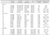
Swine, chicken, or cattle farms were studied from 2012 to 2015. Three pig farms were evaluated in 2012, and two farms each were evaluated in 2014 and 2015. Five and seven chicken farms were evaluated in 2013 and 2015, respectively, while five cattle sheds were evaluated in 2015. Participating farms were selected from the list of candidate farms prepared with help from the swine, chicken, or Korean beef cattle farmer's cooperative mentioned in the Acknowledgement section. Only farms that allowed our entrance were investigated. General husbandry characteristics of each farm are described in Table 1. Animal housing style was categorized into open type, semi-closed type, or closed type (Table 1). Open type indicates a farm in which ventilation was executed through winch curtains, while the closed type indicated farms in which a controlled mechanical ventilation system was applied without windows. The semi closed type animal confinement building installed a controlled mechanical ventilation system with upper or lower side windows. Indoor air sampling for microbial analysis or dust quantitation was performed from June to August, except for five swine farms in 2012 or 2014 (Table 1).
Indoor air sampling
To determine qualitative microbial analysis, air samples were collected at 1 to 1.5 m from the ground using a cascade impactor (BioStage; SKC, USA) at a flow rate of 28.3 L/min for 20 min in both the morning and afternoon. Since no internationally or Korean recognized guidelines regarding sampling flow rate or time or media for indoor air sampling in animal farms is available, we adopted the method described by the Korean indoor air quality test guideline of the Ministry of Environment [24] and Sampling Guide for Air Contaminants in the Workplace [10], with slight modification. Bacteria and fungi were collected in Petri dishes on different standard culture media (trypticase soy agar (TSA) and blood agar (BA) for total and pathogenic bacteria, respectively). Sabouraud dextrose agar (SDA) was used for fungi. One Petri dish for each culture media was used at each sampling time (morning and afternoon).
Indoor dust collection and endotoxin measurement
Concentration of total dust in indoor air of the animal farms was evaluated using PVC membrane filters (SKC) with 37 mm cassettes at a flow rate of 2.0 L/min for 8 h. Dust samples were usually collected from two different locations (1/3 and 2/3 distance from the exit) at each farm. The method for determination of endotoxin concentrations in the dust has been described elsewhere [32]. Endotoxins were extracted from the filters by adding 3 mL endotoxin-free limulus amebocyte lysate (LAL) water (LAL Kinetic-QCL set; Lonza, USA) with 5% Tween 20 and then shaking for one hour at 40 × g. Endotoxin concentrations from the supernatants were evaluated using the Epoch Microplate Spectrophotometer (BioTek, USA) according to the manufacturer's instructions, and were then converted into endotoxin units (EU) per cubic meter of air.
Microbial culture and identification of species
The culture plates were transported to Seoul National University, College of Veterinary Medicine for microbial cultivation and species identification. The TSA and BA plates were incubated at 37℃ for 15 to18 h. The SDA plates were incubated at 30℃ for 24 h. After incubation, colonies with different morphology, shape, smell or color were individually re-cultured in BA or SDA plates. Following simple biochemical tests including Gram staining, oxidase test, and catalase test, microbial identification was conducted using a VITEK2 automated microbial identification system according to the manufacturer's instructions (bioMérieux, France). The colonies from SDA plates were also re-cultured and identified based on colony morphology, as well as hyphae and spore characteristics after methylene blue or lactophenol blue staining.
Statistical analyses
SigmaStat 3.5 (Systat Software, USA) was used to calculate the means and standard deviations of endotoxin concentrations, as well as to identify significant differences in the endotoxin level among pig, chicken, and cattle farms. The criterion for statistical significance was set up at p < 0.05.
Results
Husbandry conditions among animal farms investigated
Evaluation of the ventilation systems for animal confinement buildings revealed that most pig farms (5 of 7) had semi-closed type animal housing in which a controlled mechanical ventilation system was primarily utilized in conjunction with natural ventilation through upper or lower side windows (Table 1). The chicken farms consisted of open (6), semi-closed (2), and closed type (4) ventilation systems. Regarding the cattle sheds, all had open type animal housing and all utilized natural ventilation supported with mechanical fans. The stocking densities of pig or cattle farms investigated were all within the recommended stocking density guide of the Korean government (0.8 m2/head for over 60 kg fattening pigs; 0.45 m2/head for 30–60 kg rearing pigs; 7 m2/head for beef cattle). The majority of broiler chicken farms investigated (8 of 10) were below the recommended stocking density guideline of the Korean government (0.066 m2/head for broilers; 0.042 m2/head for laying hens) [23]. Endotoxin levels in the total dust collected from inside the animal confinement building were highest in chicken farms (588.8 ±138.1 EU/m3) and lowest in cattle farms (57.0 ±32.1 EU/m3) (Table 1), with those of pig farms falling in between (324 ±117.6 EU/m3).
Microorganisms identified in air samples of different animal husbandry buildings
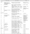
A total of six Gram-negative bacterial species, 31 Gram-positive bacterial species, and 11 fungal species were identified. Most microorganisms identified in the air of indoor livestock buildings, regardless of housing style, were Gram-positive bacteria, particularly S. lentus, S. chromogenes, Bacillus (B.) cereus, B. licheniformis and E. faecalis. These bacterial species were identified in all three animal species investigated (Table 2).
In pig farms, a total of 18 Gram-positive bacterial species, four Gram-negative bacterial species and four fungal species were identified. The most prevalent Gram-positive bacteria were S. aureus, S. cohnii spp. urealyticus and S. intermedius. In addition, Gemella morbillorum and Granulicatella elegans were isolated from two farms was and were not identified in any chicken farms or cattle sheds. Among Gram-negative bacterial and fungal species, Sphingomonas paucimobilis, and Prototheca zofii and Trichosporon mucoides, respectively, were identified in air samples from all semi-closed farms evaluated in 2012 (Table 2).
In chicken farms, a total 22 Gram-positive bacterial species, three Gram-negative bacterial species and five fungal species were identified (Table 2). The prevalent Gram-positive bacteria in farms were S. lentus, S. chromogenes, Kocuria varians, B. cereus, B. licheniformis, and E. faecalis. Among Gram-negative bacteria, Sphingomas paucimobilis was prevalent in 2013, but was identified in only one chicken farm in 2015 (Table 2). Klebsiella pneumoniae was only identified from two farms in 2015. For fungal species, Cryptococcus neoformans was isolated from four farms in 2013, but not from any farms in 2015, whereas Candida albicans was isolated from six farms in 2015 but from only one farm in 2013.
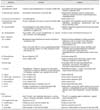
All cattle sheds evaluated in this study had similar housing style and ventilation system. Moreover, almost all farms contained Gram-positive bacteria such as B. cereus, B. licheniformis, E. faecalis, S. chromogenes and S. lentus. Only one species of Gram-negative bacteria, Acinetobacter lwoffi, was isolated (2 out of 5 farms), while Candida albicans was identified in all farms (Table 2). Major infectious diseases associated with the isolated microorganisms are briefly described in Table 3.
Isolation of microorganisms was distinct in Gram-negative bacteria. Among the three pig farms from which no Gram-negative bacteria were isolated, two farms had a closed type husbandry environment and depended solely on a controlled mechanical ventilation system. Gram-negative bacteria were only isolated from one of the four closed type chicken farms. Furthermore, endotoxin levels in the indoor dust were not significantly correlated with isolation of Gram-negative bacteria from the indoor air of the animal farms. Endotoxin levels in the animal farms from which Gram-negative bacteria were isolated were 293.9 ±87.5 EU/m3, 441.8 ±185.1 EU/m3, and 22.0 ±12.2 EU/m3 for the pig farms, chicken farms and cattle farms, respectively (Table 1). Endotoxin levels were 883.9 ±203.5 EU/m3, 168.4 ±83.4 EU/m3, and 80.3 ±52.0 EU/m3 for the pig farms, chicken farms, and the cattle farms from which Gram-negative bacteria were not isolated, respectively (Table 1).
Discussion
Livestock indoor environments may contain numerous contaminants such as microorganisms that can negatively affect the health of farm workers and animals [622]. Microbial air quality is influenced by factors such as stocking density, barn hygiene, microclimate parameters (temperature, relative humidity, gases) and ventilation system. Since the present study did not measure the microbial load, and instead only identified the microorganisms present in the air of livestock farms, we can only compare the diversity of microbial species identified in all farms with regards to previously mentioned husbandry factors that influence microbial air quality. In this study, the stocking densities of all farms except the broiler farms were within the Korean government recommended stocking density guidelines [23], and stocking density apparently had no effect on the number of microbial species isolated from the farm. All broiler farms exceeded the recommended stocking density (0.066 m2/head), which may have led to the higher endotoxin concentrations in indoor dust from chicken farms than pig or cattle farms [32]. Poultry husbandry has been reported to generate more organic dust than pig or cattle production [35].
Even though ventilation systems in the animal confinement buildings do not seem to greatly influence Gram-positive bacteria or fungi from indoor air collected from the farms, Gram-negative bacteria were apparently less isolated from the closed type farms with controlled mechanical ventilation systems than from the semi-closed or the open type farms. Gram-negative bacteria are known to be vulnerable to environmental conditions such as dehydration or radiation [29]. If the closed-type ventilation system was resulting in less humidity and/or more radiation through illumination, the indoor animal confinement building could generate more hostile conditions for Gram-negative bacteria. However, since we found no significant relationship between indoor dust endotoxin levels and the isolation of Gram-negative bacteria from indoor air, evaluation of indoor endotoxin levels might be more critical for risk assessment of animal or worker health. Even though no internationally accepted exposure limit values have been established environmentally or occupationally, airborne endotoxin concentrations over 100 EU/m3 have been reported to initiate pathophysiological conditions [182732]. Considering our present results showing endotoxin levels over 100 EU/m3 in most pig and chicken farms and previous reports [1832], it is worth preparing guidelines for occupational exposure limits or a governmental strategy for endotoxin exposure prevention.
Gram-positive bacterial species were more prevalent than Gram-negative bacterial species and fungal species. Our results agreed with those of previous reports that have identified more Gram-positive bacteria in livestock farms [6], which could again be explained by the resistance of Gram-positive bacteria to environmental conditions. Regarding the Gram-negative bacteria isolated in the present study, most of these bacteria are soil saphrophytes that have not been associated with major livestock diseases; nevertheless, they are still an important because of their ability to release endotoxins even after degradation. Exposure to high levels of endotoxins has been reported to influence immune responses towards Th2-type immune response [1832]. In addition, Gram-negative bacteria are commonly associated with hospital acquired infections in humans [11262842], indicating greater risk to immune compromised subjects. It is also important to note that the majority of microorganisms identified from most farms are linked to nosocomial infections in humans that usually involve antibiotic-resistant microorganisms. This could be the result of use of antibiotics on farm animals in the form of treatments, feed additives or prophylaxis [36], or a case of zooanthroponosis in which humans could transmit pathogens to animals. Accordingly, the antibiotic resistance of bacterial isolates should be evaluated in future studies. All fungal species reported in the present study are important pathogens to both humans and animals. Based on the rapid growth of intensive livestock production, proximity of livestock farms to communities, and easy movement of animals and humans, the threat of transmitting pathogens from animals to humans or vice versa is an important emerging public health issue.
In conclusion, the present study is believed to be the first to identify viable bacteria and fungal species in indoor air collected from all three major economic animal confinement buildings (swine, chicken, and cattle farms) nationwide in Korea. Even though the number of farms investigated for the present study was quite a small, the results could reflect overall husbandry situations in Korea since the study proceeded with help from the biggest swine, chicken, or Korean beef cattle farmer's cooperatives in Korea. Prevalent Gram-positive bacteria included Staphylococcus lentus, S. chromogenes, Bacillus cereus, B. licheniformis, and E. faecalis, regardless of the ventilation system used. Among fungi and Gram-negative bacteria, Candida albicans and Sphingomonas paucimobilis were frequently identified, respectively. All of these organisms have been reported as dangerous pathogens, especially to immune compromised or unhealthy animals or humans. It is also worth noting that quantitative evaluation of the indoor endotoxin level rather than qualitative characterization of Gram-negative bacteria could be more valuable for evaluation of organic dust-mediated health conditions in animals and animal farm workers.
 XML Download