

 PDF
PDF ePub
ePub Citation
 Print
Print


Abstract
The purpose of this study was to identify the biomechanical factors that correlate with plantar fasciitis in non-obese patients whose body mass index were below 25 kg/m2. The subjects were non-obese patients who were diagnosed as plantar fasciitis by clinical appearance, physical examination, and ultrasonographic findings (n=48), and non-obese control persons without clinical diagnosis of plantar fasciitis (n=30). The two groups were compared on fat pad thickness, ankle dorsiflexion range of motion (ROM), resting calcaneal stance position (RCSP), incidence of calcaneal spur, and calcaneal pitch. The results showed that, there were statistically significant differences between two groups in ankle dorsiflexion ROM, RCSP, and calcaneal pitch (p<0.05). Multiple logistic regression analysis showed ankle dorsiflexion ROM and RCSP strongly correlated with presence of plantar fasciitis as independent predictors (p <0.05). In conclusion, reduced ankle dorsiflexion ROM and negative RCSP (valgus tendency in rear foot) may be the biomechanical factors associated with plantar fasciitis in non-obese patients.
References
1. Crawford F, Atkins D, Edwards J. Interventions for treating plantar heel pain. Cochrane Database Syst Rev. 2000; 3:CD000416.

2. Pfeffer G, Bacchetti P, Deland J, et al. Comparison of custom and prefabricated orthoses in the initial treatment of proximal plantar fasciitis. Foot Ankle Int. 1999; 20:214–21.
3. Riddle DL, Pulisic M, Sparrow K. Impact of demographic and impairment-related variables on disability associated with plantar fasciitis. Foot Ankle Int. 2004; 25:311–7.
4. Buchbinder R. Clinical practice. Plantar fasciitis. N Engl J Med. 2004; 350:2159–66.
5. Allen RH, Gross MT. Toe flexors strength and passive extension range of motion of the first metatarsophalangeal joint in individuals with plantar fasciitis. J Orthop Sports Phys Ther. 2003; 33:468–78.
6. Irving DB, Cook JL, Menz HB. Factors associated with chronic plantar heel pain: a systematic review. J Sci Med Sport. 2006; 9:11–22.
7. Riddle DL, Pulisic M, Pidcoe P, Johnson RE. Risk factors for Plantar fasciitis: a matched case-control study. J Bone Joint Surg Am. 2003; 85:872–7.
8. Prichasuk S, Subhadrabandhu T. The relationship of pes planus and calcaneal spur to plantar heel pain. Clin Orthop Relat Res. 1994; 306:192–6.
9. Ozdemir H, Yilmaz E, Murat A, Karakurt L, Poyraz AK, Ogur E. Sonographic evaluation of plantar fasciitis and relation to body mass index. Eur J Radiol. 2005; 54:443–7.
10. Rano JA, Fallat LM, Savoy-Moore RT. Correlation of heel pain with body mass index and other characteristics of heel pain. J Foot Ankle Surg. 2001; 40:351–6.
11. Yoon K, Kim SB, Park JS. Ultrasonographic findings in plantar fasciitis. J Korean Acad Rehabil Med. 2002; 26:181–6.
12. Uzel M, Cetinus E, Bilgic E, Ekerbicer H, Karaoguz A. Comparison of ultrasonography and radiography in assessment of the heel pad compressibility index of patients with plantar heel pain syndrome. Measurement of the fat pad in plantar heel pain syndrome. Joint Bone Spine. 2006; 73:196–9.
13. Root ML, Orien WP, Weed JH, Hughes RJ. Biomechanical examination of the foot. Los Angeles: Clinical Biomechanics;1971.
14. McPoil TG, Schuit D, Knecht HG. A comparison of three positions used to evaluate tibial varum. J Am Podiatr Med Assoc. 1988; 78:22–8.
15. Younger AS, Sawatzky B, Dryden P. Radiographic assessment of adult flatfoot. Foot Ankle Int. 2005; 26:820–5.
16. Perelman GK, Figura MA, Sandberg NS. The medial instep plantar fasciotomy. J Foot Ankle Surg. 1995; 34:447–57.
17. Brown C. A review of subcalcaneal heel pain and plantar fasciitis. Aust Fam Physician. 1996; 25:875–81. ;84–5.
18. Snider MP, Clancy WG, McBeath AA. Plantar fascia release for chronic plantar fasciitis in runners. Am J Sports Med. 1983; 11:215–9.
19. Rome K, Campbell R, Flint A, Haslock I. Heel pad thickness–a contributing factor associated with plantar heel pain in young adults. Foot Ankle Int. 2002; 23:142–7.
20. Karabay N, Toros T, Hurel C. Ultrasonographic evaluation in plantar fasciitis. J Foot Ankle Surg. 2007; 46:442–6.
21. Ozdemir H, Söyüncü Y, Ozgörgen M, Dabak K. Effects of changes in heel fat pad thickness and elasticity on heel pain. J Am Podiatr Med Assoc. 2004; 94:47–52.
22. Hill RS. Ankle equinus. Prevalence and linkage to common foot pathology. J Am Podiatr Med Assoc. 1995; 85:295–300.
23. Root MI. Biomechanical examination of the foot. J Am Podiatry Assoc. 1973; 63:28–9.
24. Wearing SC, Smeathers JE, Urry SR, Hennig EM, Hills AP. The pathomechanics of plantar fasciitis. Sports Med. 2006; 36:585–611.
25. Onuba O, Ireland J. Plantar fasciitis. Ital J Orthop Traumatol. 1986; 12:533–5.
26. Williams PL, Smibert JG, Cox R, Mitchell R, Klenerman L. Imaging study of the painful heel syndrome. Foot Ankle. 1987; 7:345–9.
27. Kwong PK, Kay D, Voner RT, White MW. Plantar fasciitis. Mechanics and pathomechanics of treatment. Clin Sports Med. 1988; 7:119–26.
28. Osborne HR, Breidahl WH, Allison GT. Critical differences in lateral X-rays with and without a diagnosis of plantar fasciitis. J Sci Med Sport. 2006; 9:231–7.
29. Li J, Muehleman C. Anatomic relationship of heel spur to surrounding soft tissues: greater variability than previously reported. Clin Anat. 2007; 20:950–5.
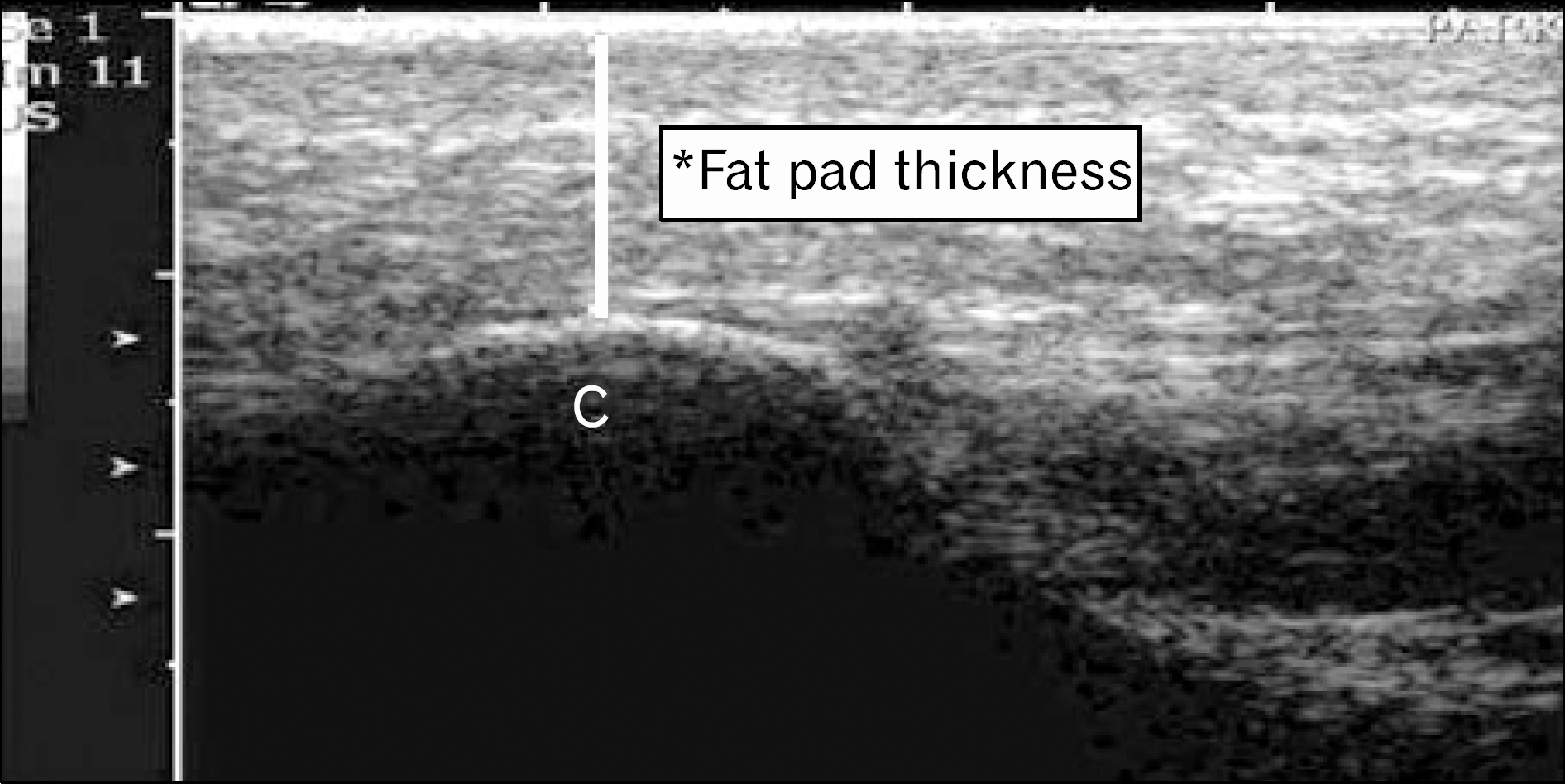
Fig. 2.
Participants were drawn a line connecting 3 points on the midline of rearfoot. And then the investigator recorded the resting calcaneal stance position with gravity goniometer.
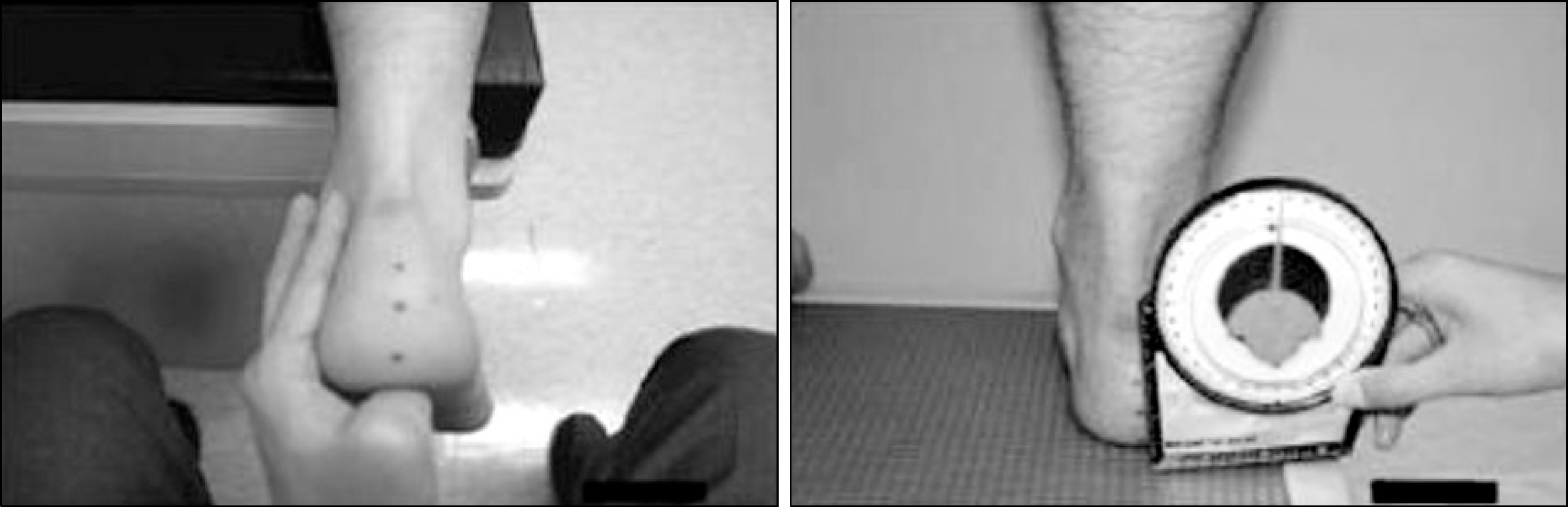
Fig. 3.
The calcaneal pitch angle, represented by a line drawn from the plantar surface of the calcaneus to the in-ferior border of the calcaneus-cuboid and line drawn from the plantar surface of the calcaneus to the inferior surface of the 5th metatarsal head.
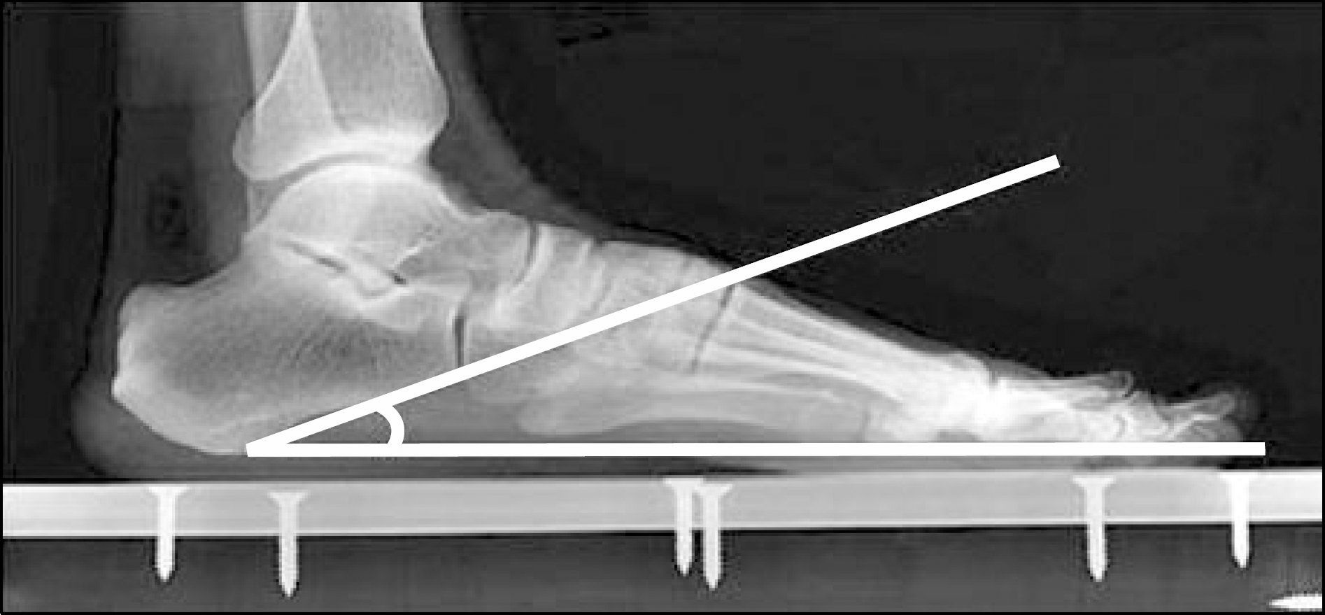
Table 1.
Demographic characteristics of subjects
| Plantar fasciitis group (n=48) | Control group (n=30) | |
|---|---|---|
| Gender | ||
| Male | 12 | 11 |
| Female | 36 | 19 |
| Age (y) | 41.6±13.2 | 38.7±13.6 |
| Body mass index (kg/m2) | x 23.8±3.7 | 24.1±3.5 |
| Affected foot Right | 27 | − |
| Left | 21 | − |
Table 2.
Comparison of biomechanical factors between plantar fasciitis group and control group
 XML Download