

 PDF
PDF ePub
ePub Citation
 Print
Print


Introduction
Accurate information of the position of impacted canine may contribute to the decision to perform a less invasive procedure when exposure of the canine is required.1 Periapical repositioning flap procedure might be a choice in labially impacted canines whereas the extensive removal of bone might be necessary for the extraction of palatally impacted canine. Also, the prognosis of an impaction can be assured accurately only when the exact position of the impacted tooth is evaluated. The use of various techniques including panoramic radiograph has been advocated for localization.2
Maxillary canine is the most frequently impacted tooth except third molar.3 Early detection of impacted maxillary canine can be possibly made in young patients, though wide variation in eruption time was reported.4 The impacted canine can be detected by combination of careful clinical and complete radiographic examinations. Clinically, digital palpation of buccal surface above primary canine root, which can reveal the position of maxillary permanent canine even in a 9- or 10-year-old patient, has been recommended as a tool for early diagnosis.5,6 Few studies considered the usage of single panoramic radiograph for the purpose. It would be advantageous to use panoramic radiographs in localizing impacted maxillary canines as it was the most commonly recommended screening radiograph, delivers relatively less radiation, easy to perform, cost effective, and readily available.7,8 The alignment of retained and displaced maxillary canines is a particular challenge in orthodontics. There are manifold reasons for this: 1. The maxillary canine has a great functional and esthetic value. 2. Following the wisdom teeth, it is the tooth most often affected by retention and displacement. 3. The orthodontic re-alignment is a difficult and extensive type of treatment.9 However, the position of this important pathological landmark has not as yet been investigated in Bangladesh. The purpose of this study was to assess the prevalence of the vertical and horizontal position of maxillary canine impaction on a basis of a single panoramic radiograph in Bangladeshi population.
Materials and Methods
The survey was a descriptive cross-sectional study to identify prevalence of impacted maxillary canines in Bangladeshi population. A random sample of seven hundred panoramic radiographs was collected from the patients' record of a dental clinic in Dhaka city. All the panoramic radiographs were taken from January 2009 to August 2010 by a single panoramic radiographic machine (Soredex, Helsinki, Finland), and the exposure time was set at 19 seconds. Among seven hundred panoramic radiographs, one hundred and twenty panoramic radiographs were excluded to minimize the selection bias. Those that were excluded from the study are: erupted and rotated canines, malaligned upper central incisor or class-II division 2 malocclusion, those patients that already had their maxillary canines extracted before 13 years of age. The study subjects included patients from 13 years to 90 years of age with the mean age 40.1 years with unerrupted maxillary canines. The remaining 580 radiographs (1160 sides) were analyzed according to the age, gender, and vertical and horizontal position of the canine impaction. On the panoramic radiograph, the height of the tip of the crown of each displaced canine was assessed in the vertical plane relative to the ipsilateral central incisor. The root of the central incisor was divided in to three zones: apical, middle, and coronal. The apical zone included the apical third of the root, and the middle zone consisted of the middle third of the root; the remainder of the root was classified as the coronal zone. For horizontal position, the following measurements were assessed. The widest mesio-distal dimension of the impacted canine was measured on a line perpendicular to the long axis. The widest mesio-distal dimension of the erupted and properly positioned canine was measured on a line perpendicular to the long axis. The widest mesio-distal dimensions of both central incisors were measured on a line perpendicular to their long axis.2,5
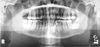
In a dim-lit room, an observer assessed each radiograph on a standard radiographic light box. Before beginning the study, the features to be evaluated for every radiograph were determined. The position of the impacted maxillary canine was recorded in line with the longitudinal axis of a tooth using the edge of a metal ruler. Data were subsequently put on SPSS 11.5 (SPSS Inc, Chicago, USA) statistical software and simple descriptive statistical analysis and chi-square (χ2) test were applied to find out the association (Fig. 1).
Results
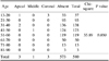
Out of 580 panoramic radiographs it was found that impacted maxillary canines were present in only 7 (1.2%) radiographs. Based on the vertical position on the panoramic radiographs, 3 (0.5%) were positioned in the apical zone, 1 (0.2%) were positioned in the middle zone and 3 (0.5%) were positioned in the coronal zone (Table 1). Most of the impacted maxillary canines that were placed coronally were found in between 13-20 years of age (Table 2). A statistical highly significant association (p=0.000) was found between the age of the patients and the vertical position of impacted canines (Table 2).
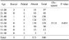
Based on the horizontal position on the panoramic radiographs, five canines (0.9%) were positioned in the buccal area, while two canines (0.3%) were placed in the palatal area (Table 3). Most of the impacted maxillary canines that were placed buccally were found in the 13-20 age group (Table 3). A statistical significant association (p=0.003) was also found between the age of the patients and the vertical position of the impacted canines (Table 4). No statistical significant association was found between the gender of the patients and the vertical and horizontal position of the impacted maxillary canine.
Discussion
The permanent canines are well recognized as important teeth, by virtue of their pivotal role in establishing the arch form, their contribution in an esthetic smile, and their participation in functional occlusion. Moreover, they are second only to the third molar as the most frequently impacted teeth. Proper localization of impacted tooth plays a fundamental role in determining the feasibility of the surgical approaches and the best access to use as well as the proper direction of application of orthodontic forces.2
The displaced maxillary canine has been the subject of many clinical studies. Our findings concurred with those studies in some respects, such as the fact that young people were significantly more often affected than the elderly.7,10 This study determined the prevalence of impacted maxillary canines in Bangladeshi population using panoramic radiographs. It has been advised that panoramic radiograph should be used with caution in performing absolute measurements or relative comparisons.11 The impacted maxillary canines were the most commonly found on the buccal side. Previous studies suggested that the percentage of buccal placement ranges from 40-75%.12-15 It varied depending on the number of cases and the technique of localization. The present study showed that 0.9% of the canines were buccally placed.
Clinical evaluation of the exact location of the impacted canine is often difficult. The clinician should, therefore, consider entirely the radiographic evidence alone.7 Several radiographic techniques for the determination of the position of the unerrupted maxillary canines have been advocated in the past either in a single radiograph or in combinations, with every effort taken towards minimizing the radiation dose and cost while maximizing the information.16
Panoramic radiograph was considered for the localization of impacted canines for a period of time, however, regardless of the method applied, the conventional radiographs could not show the exact appearance of the impacted tooth in its longitudinal axis and the relationship with the neighboring bony structures,15 even there were a few studies on localization using single panoramic radiograph.17,18 In the present study it was found that in the vertical plane of the impacted canine, most of the canines were present in the coronal and apical zone. In the horizontal plane most of the impacted maxillary canines were present in the buccal side. These findings were similar to the studies performed in Israeli and Indian populations.7,16
In this study it was found that most of the impacted maxillary canines were present in young people especially between 13 and 30 years of age, which was similar to a study in India.16 Adjacent anomalous or missing maxillary lateral incisors have been implicated in the etiology of palatally displaced canines by not providing proper guidance to the canine during its eruption. However, a recent review of the literature suggested that the etiology of palatally displaced canines was genetic in origin. The etiology of labially or buccally impacted canines might be different from the displaced canine, being due to inadequate arch space. Cross-sectional occlusal radiographs are recommended for localization, however they have limitations.19
A large number of impacted maxillary canines were excluded from the study because they were rotated, resulting in a projection that was the mesial or distal view of the same tooth on the panoramic radiographs. The sample size was as small as 160 subjects in the study by Chaushu et al, and the same exclusion criteria was also applied.7 The largest number of impacted canines were projected in the coronal (5) zone and buccal (5) zone. In a previous study, 66.15% of buccally impacted canines were found in the coronal zone, and 74.74% of palatally impacted canines were found in the middle (8) zone. Only three (0.5%) impacted canines were present in the apical zone, and two (0.3%) canines were present in the palatal zone. This was in contrast to 14 (8.75%) labially and 6 (3.75%) palatally impacted canines in the apical zone in the study by Chaushu et al.7 In our investigation most of the impacted canines in the apical zone were excluded as they were either rotated or horizontally placed.
In conclusion, impacted canines can be detected at an early age, and clinicians might be able to prevent them by means of proper clinical diagnosis, radiographic evaluation and timely interceptive treatment. Surgical techniques that can be used to manage impacted canines vary depending on whether the impactions are labial or palatal, and the orthodontic techniques vary according to the clinical judgment and experience. The clinician needs to be familiar with the differences in the surgical management of the labially and palatally impacted canines, the best method of attachment to the canine for orthodontic force application, the advantages of one-arch versus two-arch treatment, and the implications of canine extraction. A single panoramic radiograph can serve as a reliable indicator for determining the bucco-palatal position of the impacted permanent maxillary canines when they lie in the middle and coronal zones with respect to the ipsilateral central incisor. Prevalence was found to be low in present study because a large number of impacted maxillary canines were excluded from the study because they were rotated, resulting in a projection that was the mesial or distal view of the same tooth on the panoramic radiographs, and some patients had their maxillary canines already extracted.


 XML Download