

 PDF
PDF ePub
ePub Citation
 Print
Print


Introduction
As a result of globalization, migrations are very common now. Transnational crime has also increased as a consequence. To identify the ages of victims or suspects is a necessary task in forensic science. Age identification can be directly performed from identification cards or passports, but in cases without identification papers, refugees, or cadavers, more complicated age identification methods may be needed.
Human teeth can be used to identify chronological age. Tooth development in humans can indicate age within certain ranges.1,2 Each tooth type develops at different ages. The third molars, or wisdom teeth, are the teeth that develop and erupt last clinically. Many studies have identified various characteristics of third molar eruption with age.3-6 Some authors have claimed that the age of third molar eruption varies by ethnicity.7-9 Others have described sex differences in the age of eruption, with some contradictory findings.4,5,10
This study focused on the level of third molar eruption for age determination; only three previous studies have focused primarily on this question.1-3 Furthermore, all of those studies examined only vertically erupting teeth.
This study, thus, aimed to determine the relationship between the stage of third molar tooth eruption (both vertical and mesio-angular) and chronological age.
Materials and Methods
Subject recruitment and image preparation
This retrospective study examined the panoramic radiographs of 701 Thais taken in 2008-2009 from the records of the Oral Radiology Clinic of the Faculty of Dentistry, Chiang Mai University, Chiang Mai. All of the panoramic radiographs were taken using the same panoramic X-ray machine (Cranex Tome, Soredex, Helsinki, Finland). The age of the subjects ranged from 10 to 30 years. A total of 1,993 third molars were found on the panoramic radiographs. The exclusion criteria were 1) no adjacent second permanent molars, 2) overlapping of the second and third molar, 3) having a history of endocrine gland diseases or syndromes such as gigantism or rickets that might disturb growth and development, 4) presence of an eruptional axis other than vertical or mesio-angular, and 5) presence of a fixed orthodontic appliance.
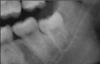
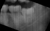
The data records were examined for sex, date of birth, and medical history. Each panoramic radiograph was photographed by camera and stored in JPEG files before being shown to the observers. This study used ImageJ software version 1.4 (NIH, Bethesda, USA) to prepare the images before data recording. The region of interest, which covered the entire second molar and third molar, was cropped to a size of 300×250 pixels and converted to 8-bit gray scale. Next, the first reference point was located at the dentinoenamel junction (DEJ) of the distal side of the adjacent second molar tooth (Fig. 1). The "grid" function in the software then was applied to each image. This study used a grid size of 100 pixel squares per grid space (Fig. 2). The image was rotated until the occlusal plane of the second molar was parallel to one of the horizontal lines of the grid.
Observer calibration
Two maxillofacial radiologists with more than 10 years' working experience were the observers in this study. To calibrate them, both practiced scoring tooth height using the grid and following the distance measurement instructions until their results were comparable. Their kappa analysis score was 0.85 at a significance level of p=0.01.
Observation strategy
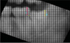
The grid square in which the DEJ was located was established as level 1, with each more occlusal square assigned successively higher numbers until the occlusal plane was reached.
We scored the position of the most occlusal point of the third molar crown by the number of complete grid squares from the level of the second molar DEJ, rounding up or down. Squares below the second molar DEJ were given negative numbers (Fig. 3). The distance ratio (D) was calculated as the number of complete grid squares from the DEJ of the second molar to the most occlusal point of the third molar divided by the number of complete grid squares from the DEJ of the second molar to its occlusal plane. The ratio results then were categorized into four ranges: 1) D<0; 2) 0≤D<0.5; 3) 0.5≤D<1; 4) D≥1.
However, negative values of D would have given spurious results when correlation analysis was applied. A mechanism was devised to overcome this problem. The greatest distance between the occlusal plane of the second molar and the most occlusal point of the third molar was 18 grid spaces. Nine grid spaces were above and nine below the DEJ of the second molar. The DEJ level was given a score of 10 to allow us to use 18 as the maximum distance score. Later, the height ratio (H) was calculated from the score of the third molar divided by that of the adjacent second molar. Correlations then were analyzed between those H values and the ages of the samples in both females and males, using the Pearson correlation coefficient calculated with SPSS version 16.0 (SPSS Inc., Chicago, IL, USA), with p<0.05 considered significant.
Results
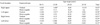
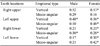
There were 701 panoramic radiographs with 1,993 third molars with various eruptional axes in this study. The age of this population ranged from 10 to 30 years. There were 738 third molars in males and 1,255 in females. Among them, 1,708 impactions were vertical or mesio-angular. The distribution of the samples in terms of age range, sex, and tooth location are shown in Table 1.
Table 2 presents the mean age classified by ranges of D in the vertically erupted third molars. The mean ages of the samples who had an upper vertical erupted third molar at or above the DEJ but below one half of the coronal height of the adjacent second molar were 20.1 years and 19.8 years on the right and left sides in the males, and 19.9 years and 20.2 years in the females, respectively. Furthermore, in the case of the lower molars, the patients' mean ages were 21.8 and 20.8 years on the right and left sides in the males and 21.0 years and 20.3 years in the females, respectively. Table 3 also presents the mean age classified by ranges of D for the mesio-angularly erupted teeth.
The upper third molar teeth with vertical eruption presented a medium level of correlation between the H and age with statistical significance (p<0.05). The age range from 15 to under 20 years showed a medium level of correlation between the height ratio and age at p<0.05 (Table 4). Moreover, the results also demonstrate that there was a medium level of correlation between both sexes and the height ratio of the upper third molar teeth, especially in teeth with vertical eruption (Table 5).
Discussion
Age identification is necessary for some legal reasons. Several methods of age identification have been proposed. In the past, some studies showed good reliability in age predictions from third molars.11,12 However, they mainly focused on root calcification and age. Our study was focused on the correlation between the level of eruptive height of the third molar and age identification. This study was similar to a study by Olze et al,3 but their study classified the level of the third molar tooth into four categories: 1) same level as the adjacent second molar, 2) gingival eruption, 3) alveolar bone eruption, and 4) still underneath the bone. We found that it was sometimes difficult to identify on a panoramic radiograph if there was a gingival eruption or just a bone eruption. Therefore, using the DEJ as a reference point may be more reliable, especially when there is no remaining soft tissue, such as in postmortem cases with tissue decay because the DEJ still persists even when all soft tissue has disappeared. Moreover, as we digitized each panoramic radiograph, the image quality was enhanced and the DEJ was easily visualized and located.
We chose ImageJ software, which provides a function named "Grid", to level up the occlusal plane of the second molar teeth, facilitating the observers' visualization and data recording. This study was not designed to use linear measurements; we chose a grid instead. On panoramic radiographs, there is an overlap between lingual cusps and buccal cusps, and it was sometimes difficult to identify which cusps represented the highest points. Therefore, using one grid space as a unit of measurement allowed us to average the height of the coronal part of the tooth and was also more practical than using a ruler.
A study of age prediction from third molars by Thevissen et al13 also used the DEJ as a reference point; however, they focused on comparing the root length of second molars and third molars. Our study found that the greatest statistically significant correlation between tooth eruption and age was found in the 15 to <20 year age range, and this range is the same as the growth spurt period, so this may explain why we obtained such a result. Vertical eruption showed better correlation than mesio-angular eruption with chronological age. This is not surprising, because only bone presented an obstacle to eruption in vertical eruption, whereas in mesio-angular eruption or impaction, the obstacle was both bone and tooth, which is harder than bone. This study also found that the upper third molar showed greater correlation than the lower third molar eruption with chronological age, and we cannot explain why. Perhaps gravity or the density of the alveolar ridge can affect this, and greater numbers of mesio-angular teeth and a greater distribution of eruption angles should be investigated in further research.
The eruptional level in this study, which was at or above the DEJ of the adjacent second molar, was similar to the gingival eruption level of a study of Olze et al.2 Our results showed a mean age of 20.1 years in males, which is close to that of the African male group in another study by Olze et al, but in the females in our study, the mean age was 19.9, which is close to that of the German female group in the comparative study by Olze et al. The females in our study seemed to have earlier tooth eruption than the males.
Another ratio which may be of benefit for age prediction is the ratio representing a level of third molar eruption which is between the middle of the second molar crown height and its occlusal surface. Using this ratio, in both males and females, our results show a mean age of around 22 years. The ratio representing a level of third molar eruption which is at the level of the adjacent second molar or above, may not be able to provide meaningful data for age identification. Normally, vertically positioned teeth erupt until they reach the occlusal plane if there is no obstacle and then stop their eruption, or stop at a higher level than the occlusal plane if there is no opposing tooth. However, some teeth in this study may have reached the occlusal plane before we produced radiographs. In such cases, the age identification may not be reliable.


 XML Download