

 PDF
PDF ePub
ePub Citation
 Print
Print


Ovarian vein thrombosis (OVT) is a rare cause of acute abdominal pain, and it affects approximately 1 in 2,000 deliveries or abortion [1]. However, this diagnosis should be considered not only in postpartum patients but also in women with pelvic inflammatory disease (PID), with malignancy, who went through recent abdominal surgery, or has known hypercoagulable state [2]. Common symptoms of OVT are fever, nausea, vomiting, chills, lower abdominal pain, tachycardia, and flank pain. The clinical manifestations are variable from asymptomatic and dull abdominal pain to sepsis, pulmonary embolism and life threatening events. The complications of OVT can be significant, so careful diagnosis, which is based on computed tomography (CT), color Doppler ultrasonography and magnetic resonance imaging (MRI), is important [3]. Differential diagnosis must be included appendicitis, peritonitis, adnexal torsion, tubo-ovarian abscess, ectopic pregnancy, intussusception, and pyelonephritis [1]. We report a case of a patient presenting with OVT which is accompanied with PID, and review the diagnostic and therapeutic options for this disease.
Case Report
A 27-year-old healthy nulliparous woman was visited Gangnam Severance Hospital emergency department with one day of aggravating right lower quadrant pain. The past medical history and review of systems were not remarkable. There was no family history of hypercoagulable state. Transvaginal ultrasound was within normal range. The abdomino-pelvic CT revealed mild pelvic ascites with smooth peritoneal thickening which is suggesting PID. She was admitted to gynecologic department under the impression of PID and received an intravenous aminoglycoside and metronidazole for 8 days as a empirical antibiotic treatment. She had vaginal discharge and the result of cervical culture was positive for gardnerella and chlamydia. The patient was discharged after 8 days hospital stay with mild improvement in her symptom. She was to continue 1 week of oral aminoglycoside and metronidazole.
After 5 days, she revisited emergency department for recurrent abdominal pain, which is more aggravated and localized at right lower quadrant area. On physical examination, the patient had acute ill-looking appearance and was afebrile, with normotensive and not tachycardic heart rate. Examinations revealed direct tenderness and rebound tenderness in right lower quadrant area. Moderate cervical motion tenderness and right adnexal tenderness were observed during pelvic examination. Transvaginal ultrasound was unremarkable except right hydrosalpinx. Routine laboratory findings were within normal limits. There were no leukocytosis (white cell count, 8,630) and no elevation of C-reactive protein (P<0.78).
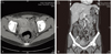
She was readmitted to gynecologic department and abdominopelvic CT, which was performed to evaluate persistent pain for differentiation of acute appendicitis and surgical abdomen, revealed the partial focal thrombosis of right ovarian vein (Fig. 1) and secondary appendicitis suggesting PID, with no other evidence of cause of her recurred pain. The diagnosis was confirmed as OVT induced by PID and anticoagulation treatment was started as intravenous unfractionated heparin infusion (normal saline 500 mL + heparin 25,000 u; rate, 40 mL/hr). After four days of this therapy, her pain was resolved. She discharged on a regimen of warfarin (5 mg daily) and this regimen was maintained for 6 months. Follow-up abdomino-pelvic CT after 3 months revealed complete resolution of OVT. She has no further complications through 13 months of follow-up.
Discussion
OVT is very rare cause of acute abdominal pain and it is diagnosed predominantly after delivery or abortion, especially during the early postnatal period, and is seldom idiopathic [1,2]. OVT is developed 0.05% to 0.18% of deliveries [3,4]. In one study, OVT is affected in 1 in 200 cases of febrile abortion [5]. The incidence outside the postpartum period is not evaluated, but it can be associated with PID, recent gynecological surgery, underlying malignancy, and hypercoagulable state. Antiphospholipid antibody syndrome (APS) is also considerable disease which is characterized by recurrent vascular thrombosis, pregnancy morbidity [6]. But Screeing study for APS was not performed in our case because this was her first thrombotic event and was not related with pregnancy. PID was the only risk factor in our patient.
Most of the cases of OVT, about 70% to 90%, develop in the right ovarian vein, so these patients are easily misdiagnosed as acute appendicitis [3]. This might be due to the physiologic dextrorotation of the pregnant uterus towards the right and the longer right ovarian vein with less competent valve. It causes blood flow stasis and at term, the diameter of the ovarian vein increases to 3 times larger than that of ovarian vein before pregnancy. Blood flow in the ovarian vein rapidly decreases right after delivery [5]. The OVT, which is not associated with pregnancy, also is predominantly occurred in the right ovarian vein [3,7]. Ovarian veins have enormous anastomosis with the uterine and vaginal venous plexuses, thus it is the cause of a portal of entry for bacterial seeding [8]. This retrograde drainage pattern from the left ovarian vein and anterograde flow into the right ovarian vein, especially in the presence of PID, may expose the right ovarian vein to an infectious environment [5].
Early diagnosis of ovarian vein thrombosis is essential to prevent life-threatening sequelae.
OVT may be diagnosed with modern imaging techniques, such as ultrasound, CT, or MRI [3]. Ultrasound is the most convenient imaging modality when postpartum complications are suspected. Findings of ultrasound are an anechoic to hypoechoic mass between the adnexa and the absence of blood flow within the mass. However, sonographic images can be limited, and the sensitivity of Doppler ultrasonography is known as about 50% [3]. A suspicious ultrasound should be followed by CT or MRI. CT imaging presents a tubular mass between the adnexa. Contrast-enhanced images of the ovarian vein, which are critical in establishing the diagnosis, reveal a low-density lumen with sharply defined walls. Contrast-enhanced CT is currently considered the diagnostic imaging modality of choice. The sensitivity of CT scan is reported from 95% to 100% [9]. The OVT findings from MRI is a tubular mass with low signal intensity on T1-weighted images and moderate signal intensity on T2-weighted images. In one study, the sensitivities and specificities for MRI were 92% and 100%, respectively [3]. The accuracy of these imaging techniques was not currently confirmed, thus the best method to diagnose OVT remains controversial.
Main treatment of OVT is broad-spectrum antibiotics and anticoagulation. Intravenous antibiotics such as clindamycin and gentamicin, imipenem and ampicillin and sulbactam, or single-agent therapy with a second- or third-generation cephalosporin are proper to treat this diagnosis. Anticoagulation such as heparin has also been administered to shrink small septic emboli and hasten fever resolution. Treatment regimens consists of 5,000 U i.v. bolus heparin followed by an initial maintenance dose of 1,000 U/hr or 10,000 units daily (5,000 U administered b.i.d.) [10]. The goal to obtain is an activated partial thromboplastin time of 1.5 to 2 times normal, and the titration rate of heparin should be modified for therapeutic target [2]. There are no standard protocols for duration of antibiotics and anticoagulation therapy in OVT. Usually recommended period is 3 to 6 months anticoagulation treatment and 7 to 10 days course of antibiotics in reported cases [1,3,4]. In our patients, repeat CT scan which was performed at 3 months after diagnosis revealed resolution of thrombus, but maintaining of warfarin therapy for 3 more months was required because of persistent mild lower abdominal pain.
Heparin has a number of side effects including bleeding, heparin-induced thrombocytopenia and osteoporosis. The low-molecular-weight heparins (LMWHs) have many advantages over unfractionated heparin [10]. LMWH have replaced traditional heparin therapy for both prophylaxis and treatment of thromboembolism. Although no significant difference between two groups was observed in venous thromboembolism, the LMWH therapy was reported with a reduction in major bleeding and heparin-induced thrombocytopenia and osteoporosis as well [2,10]. However there is few data to support their efficacy in OVT [2]. In our case, we used only IV unfractionated heparin and oral warfarin. Further investigations are required to determine whether conventional unfractionated heparin or LMWH in particular is beneficial in OVT. Surgery is indicated in some cases after the failure of medical treatment or whenever the risk of pulmonary embolism is high. In fact, the risk of pulmonary embolism is seen in 13 to 33% and a mortality of pulmonary embolism was up to 4% [11]. Thus, the aim of surgery is to prevent this high embolic risk in those cases that are not responsive to medical treatment.
In conclusion, awareness of this diagnosis and clinical suspect in a patient suffering from lower quadrant pain and fever, which does not respond to adequate antibiotics, is important in appropriate diagnosis. However, the diagnosis OVT is still difficult these days [12].
 XML Download