

 PDF
PDF ePub
ePub Citation
 Print
Print


INTRODUCTION
Anatomical reduction and stable internal fixation have been shown to be most critical factors in acetabular fracture management1,2). To obtain anatomical reduction with the lowest incidence of complications, it is essential to understand the plane of displacement in fractured fragments and the main fragments, which are not displaced. This might be of great value when choosing the correct reduction maneuver in each individual fracture types. In both transverse and both column acetabular fractures, it has been suggested that the head of the femur is displaced medially with the ischiopubic fracture segment3,4). We hypothesized that the central dislocation of the femoral head does generally not occur in transverse acetabular fractures, although it does usually occur in both column fractures. To demonstrate this hypothesis, we analyzed the measurements of femoral head and superior iliac segment displacements as well as the ischiadic fragment rotation on two-dimentional plain radiographs.
MATERIALS AND METHODS
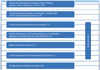
This retrospective study included records of all patients admitted to Kocaeli University Hospital in Turkey with acetabular fracture between January 1, 2001 and March 31, 2014. Overall, 276 patients were identified. Of these, 75 patients had transverse (simple transverse and transverse-posterior wall) or both column acetabular fractures. Different orthopaedic trauma surgeons reviewed and classified preoperative plain radiographs and computed tomography (CT) images of the patients. Acetabular fractures were classified according to Letournel-Judet classification system3). Twenty-three patients were excluded from the study, of these eleven patients had conservative treatment. Ten patients had disruption of the pubic symphysis and contralateral displaced associated pelvic injury causing ischiopubic segment rotation in multiple planes, which might affect the measurements. Additionally, one patient who had bilateral acetabular fracture and the other one who had X-Ray images that did not include sacroiliac joints were also excluded (Fig. 1). The remaining 52 patients who had been operated by two authors were included in the study. Twenty-five patients had transverse and 27 patients had both column acetabular fractures.
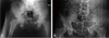
Superior iliac segment and inferior ischiopubic segment (femoral head) displacements were measured based on preoperative plain anteroposterior (AP) view radiographs of all patients (Fig. 2). The AP view radiographs were taken after the reduction of dislocated hip joint under sedation in 17 patients, and the measurements were made on these radiographs. The vertical axis of the pelvis (VA line) was defined by connecting the middle of the inter-sacroiliac line and the middle of the pubic symphysis in preoperative AP view radiographs5). Two perpendicular lines were drawn to the VA line across top points of greater sciatic notch. The distances (a) between the fracture side sciatic notch and the VA line and (b) between the contralateral intact sciatic notch and the VA line were measured. The a/b ratio corresponded to the superior iliac segment displacement or rotation. Likewise, two perpendicular lines were drawn to the VA line across each medial border of femoral head. The distances (c) between the fracture side femoral head and the VA line and (d) between the contralateral intact femoral head and the VA line were measured. The c/d ratio corresponded to the femoral head displacement. The c/d ratio that was smaller than one showed the central dislocation of femoral head. The width of ischium was measured (e) on fractured side and (f) contralateral side. The width of the ischium increased as internal rotation of the ischiadic fragment increased6). The e/f ratio increment reflected ischiadic fragment mobility. Two authors (OS and AYS) performed the measurements independently and the mean values were used in statistical analysis. The interobserver reliability was evaluated with intraclass correlation coefficient (ICC). ICC values less than 0.5, between 0.5 and 0.75, between 0.75 and 0.9, and greater than 0.90 are indicative of poor, moderate, good, and excellent reliability, respectively.
Associated posterior pelvic injuries were evaluated from CTs of all patients. Posterior pelvic injuries including sacral fractures or sacroiliac joint separations were recorded. The postoperative acetabular reduction was assessed according to Matta's classification7). The largest residual displacement was recorded in millimeters on postoperative plain radiographic views. Displacements of 1 mm or less were considered as anatomic, 2 to 3 mm as imperfect, and greater than 3 mm as poor reduction7). Comparative studies were performed between groups, which were formed according to acetabular fracture type, a/b ratio, c/d ratio, associated ipsilateral posterior pelvic injury, and postoperative reduction quality.
Statistical analysis of the data obtained from 52 patients was performed using IBM SPSS Statistics for Windows, version 20.0 (IBM Co., Armonk, NY, USA). Normality of data was ascertained using the Kolmogorov-Smirnov's test. The quantitative data of a/b was demonstrated as mean±standard deviation and compared across different types of fracture and reduction quality using Student t-test. The median values were calculated for discrete variables (c/d and e/f ratio). The Mann-Whitney U test was used to evaluate the statistical significance of discrete variables. Spearman's correlation coefficient was used to analyze whether significant correlation exists between the discrete variables. The categorical factors were analyzed using Yates chi-square test and Fisher's exact tests. A P-value of less than 0.05 was considered statistically significant. The power analysis was performed using the G*Power 3.1.9.2 (Franz Faul, Universitat Kiel, Germany).
RESULTS
In our study, 39 of 52 patients were male (75.0%). The mean age was 39.6 years (range, 18-61 years). The most common injury mechanism was traffic accidents (42 patients, 80.8%) followed by falling from height (10 patients, 19.2%).
The superior iliac fragment displacement (a/b) range of the transverse fracture group ranged from 0.92 to 1.22 with a mean of 1.06±0.09 (Table 1). The median value of femoral head displacement (c/d) of the transverse fracture group was 1.02 (1.000-1.07). Twenty-two of 25 transverse fractures have c/d ratio ≥1. Ischiadic fragment rotation (e/f ratio) of the transverse fracture group was 1.000 (1.000-1.000). The superior iliac fragment displacement (a/b) range of the both column fracture group was 0.83 to 1.12 with a mean of 0.98±0.07 (Table 1). The median value of femoral head displacement (c/d) of the both column fractures was 0.78 (0.64-0.85). All fractures of both column group had c/d ratio <1. Ischiadic fragment rotation (e/f ratio) of the both column group was 1.15 (1.06-1.23). The difference between groups in a/b, c/d, and e/f ratio were statistically significant (P=0.003, <0.001, and <0.001, respectively). A negative correlation emerged between femoral head displacement (c/d) and ischiadic fragment rotation (e/f) (P<0.001, r=–0.697).
In 10 (40.0%) of the 25 transverse fractures, associated ipsilateral posterior pelvic injury was also present. Among these patients, 9 were sacroiliac separations and one was sacrum fracture. Only 3 (11.1%) ipsilateral posterior pelvic injuries were detected in 27 both column fractures. The contralateral displaced pelvic injuries were excluded from the study, and for this reason, only ipsilateral posterior pelvic injuries were assessed statistically. Ipsilateral posterior pelvic injuries were significantly more common in patients with transverse than both column acetabular fractures (P=0.016).
The reduction quality of transverse fractures (n=25) was graded radiographically, according to Matta's criteria, as anatomical in 11 (44.0%) cases, imperfect in 8 (32.0%) cases, and poor in 6 (24.0%) cases. Acetabular reduction was anatomic in 10 (37.0%) cases, imperfect in 10 (37.0%) cases, and poor in 7 (25.9%) cases among the both column fractures (n=27). When patients were grouped according to reduction quality, no significant difference in a/b and c/d ratio emerged between the groups (P>0.05).
A high interobserver reliability of a/b, c/d, and e/f was evaluated using ICC, yielding the values of 0.71, 0.89, and 0.78, respectively. Power analysis demonstrated that the power of the study was 0.997 (α=0.05).
DISCUSSION
The transverse fracture line divides the acetabulum into two segments, which are superior iliac and inferior ischiopubic fragments3). Three main fragments identified in both column fractures are the posterior iliac fragment, iliopubic fragment (anterior column), and ischiadic fragment (posterior column)8). Judet et al.3) suggested that the main displacement in transverse and both column acetabular fractures occurs in the ischiopubic fragment with central dislocation of femoral head. Recently, Pierannunzii et al.8) suggested that in both column acetabular fractures iliopubic and ishiadic, fragments are pushed medially by the femoral head with a central dislocation in all instances. The main problem is that no one objective method exists to measure the degree of femoral head displacement to be described in acetabular fractures. Dickson and Matta6) showed that the width of ischium on the AP radiograph increased as internal rotation increased in pelvic fracture patients. Our analysis of the widths of ischiums in transverse and both column fractures showed internal rotation of ischiadic fragment in both column fractures, supporting the suggestion of Pierannunzii et al.8) and Judet et al3). However, in the transverse acetabular fracture group, the inferior ischiopubic fragment showed low mobility.
Transverse fractures are assumed to result from a lateral compression (LC) force transmitted via the trochanter, proximal femur, or axially along the femur if the hip is in a flexed position at the time of impact3). Two recent papers by Osgood et al.9) and Suzuki et al.10) analyzed the injury mechanism in combined pelvic-acetabular injury. Osgood et al.9), analyzing 40 cases, found the injury mechanism to be either AP compression (APC) or LC. The study of Suzuki et al.10) revealed 62 posterior pelvic lesions associated with transverse-type acetabular fractures, with the majority having ipsilateral sacroiliac disruption. In contrast to the previous studies, recent studies suggested that APC mechanism is just as common as the LC mechanism of injury in transverse fractures. Our study supports the potential dominance of combined injury mechanism in transverse acetabular fracture with external rotation of superior iliac fragment and ipsilateral posterior pelvic injury.
According to Bastian and Giannoudis4), central dislocation of femoral headwere reported in combination with posterior column, anterior column, transverse, T shaped, transverse and posterior wall, anterior column and posterior hemitransverse or associated both column acetabular fractures. In all these fracture types beside transverse fracture, fracture line reached to obturator foramen and femoral head was medialized. However according to our opinion, in transverse fractures femoral head was not medialized as ischiopubic fragment was intact (as there weren't any injury in obturator foramen or symphisis pubis).
The value of this study may be limited due to its retrospective design, measurement via the two dimensional imaging, and a relatively small number of patients. The use of our measurement method is limited in cases with contralateral associated pelvic injury. Further studies are needed to measure the femoral head displacement with more exact three-dimensional analysis.

 XML Download