

 PDF
PDF ePub
ePub Citation
 Print
Print


Amyloidosis is a disease caused by the accumulation of protein polysaccharide complex or insoluble fibrillar protein. This accumulation most commonly occurs in the kidneys, heart, and gastrointestinal tract, and invasion to osseous tissues is relatively uncommon1). Depending on the amino acids sequences of fibrillar protein, the disease can be classified into AA, AL, AH, ATTR, or Aβ2M. Of these, AL amyloidosis is one of the most common (found in 10-20% of clinical multiple myeloma)2). Symptoms of amyloidosis vary depending on the size and amount of deposition, amyloid arthropathy is shown in approximately 5% of systematic amyloidosis3). Although there are a few studies reporting multiple myeloma combined with amyloid arthropatthy, amyloid arthropathy involving hip joints globally45), there is no report from South Korea describing amyloid arthropathy of a hip joint treated with total hip arthroplasty. Therefore in the present report, the authors report a case of total hip arthroplasty in an amyloidosis patient and include a relevant literature review.
CASE REPORT
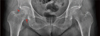
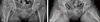
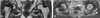
Since about a year ago visiting our hospital, A 73-year-old female patient visited our clinic with both hip pain more severe on the right side. The patient experienced pain while walking and the pain was getting more serious, she did not have a history of trauma. The patient has been taking drugs for diabetes and hypertension for the past 10 years. Approximately 8 years ago, she underwent percutaneous coronary intervention and total thyroidectomy for angina pectoris and thyroid cancer, respectively. Five years ago, the patient was also diagnosed with end stage renal disease (ESRD) and taking the required drugs. After the onset of symptoms, a simple pelvis X-ray image taken by another institute indicated osteolytic lesions on the right femoral head and neck (Fig. 1), computed tomographuy (CT) images of the pelvis also showed similar osteolytic lesions; both hip joints were swollen and the joint cavities were invaded (Fig. 2). In order to confirm the presence of lesions, non-enhanced magnetic resonance images (MRI) were taken of both hip joint and the right femoral head and neck showed osseous lesions. In both joint cavities, low signal intensity of lesion deposition were observed (Fig. 3). Approximately 1 year later, the patient was subjected to simple X-ray and enhanced MRI and we identified subchondral inflammation and that the femoral head and neck cysts were enlarged. Further, amount the lesion which shows low signal intensity in MRI that means deposition in cavities increased as well (Fig. 4, 5).
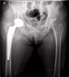
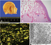
The range of motion of the right hip joint, the flexion contracture, further flexion, internal rotation, and external rotation were measured and determined to be 10°, 90°, –10°, and 40°, respectively. In addition, adduction and abduction were 50°, and 30°, respectively. To make a more accurate diagnosis, ultrasound-guided biopsy was performed and amyloidosis was confirmed based on apple-green birefringence with Congo red stain and the presence of amyloid P positive cells when immunohistochemical analysis was conducted. When hospitalized, hematological analyses were conducted; erythrocyte sedimentation rate was 110 mm/hr and C-reactive protein was 2.95 mg/dL. In addition, it was suggested that the patient may have anemia (hemoglobin 9.5 g/dL). Despite of conservative treatment, the patient still complained of serious pain, thus total hip arthroplasty of the right hip was performed to relieve pain, prevent femoral neck fracture and the enlargement of lesions (Fig. 6). While performing surgery, the authors confirmed cystic formation of the femoral neck and head and massive joint effusion, synovial hypertrophy, and amyloid deposition throughout the joint muscles (Fig. 7).
According to the biopsy, apple-green birefringence with Congo red stain and amyloid P positive cells were observed and amyloid fibrils were visible with an electron microscope (Fig. 8). No microbial contamination was observed with ultrasound-assisted biopsy and culture examination. To further test if amyloidosis invaded toward other tissues, esophagogastroduodenoscopy, sigmoidscopy, chest CT, and abdomen ultrasonic wave examinations were performed and the results suggested no additional amyloidosis. Serum and urinary protein immune electrophoresis were performed to elucidate the primary cause of amyloidosis; in this, lambda light chain (monoclonal gammopathy, lambda type) was found to be high and the bone marrow aspiration examination indicated increase in plasma cells (19.4%). Therefore, the patient was diagnosed with secondary multiple local amyloidosis due to multiple myeloma. The patient is now being treated for multiple myeloma and followed up.
DISCUSSION
Amyloidosis-a disease first described by Wilks in 1856-is characterized by the extracellular deposition of amyloid protein in various tissues and organs6). Depending upon the clinical phase, amyloidosis can be classified as (1) primary amyloidosis, (2) secondary amyloidosis, (3) local amyloidosis, (4) genetic familial amyloidosis, or (5) degenerative amyloidosis. In 1990, the International Nomenclature Committee for Amylodosis suggested to classify amyloidosis based on amino acid sequences of fibril proteins such as AA, AL, AH, ATTR, and Aβ2M.
The cause of primary amyloidosis can be classified to primary amyloidosis without other diseases and amyloidosis with multiple myeloma. Approximately 5-15% multiple myeloma patients have amyloidosis. In primary amyloidosis, immunoglobulin kappa and lambda light chain act as a precursor protein, thus it is called AL amyloidosis. Secondary amyloidosis which is caused by infectious diseases (e.g., tuberculosis, leprosy, and chronic osteomyelitis), chronic inflammatory diseases (e.g., rheumatoid arthritis, and ankylosing spondylitis), and malignant tumors (e.g., renal cell carcinoma, and Hodgkin's disease) is called AA amyloidosis. This is because serum amyloid A (SAA; an acute phase reactant) is increased in the serum.
Amyloid deposits can invade various tissues including the kidneys, heart, lung, liver, skin and joints thus it represents variable clinical patterns. The amyloid deposition site depends on the type of the fibril protein. Typically AL type protein deposits on the heart, tongue, gastro-intestinal tract, neuronal system, and skin whereas the AA type is typically found in the liver, spleen, and kidneys. The major differences between the AL and AA type includes not only the structure of precursor amyloid protein but also clinical patterns. In particular, arthropathy is known as a disease specific finding of AL type7).
Amyloid arthropathy can be divided into two types. If amyloid is deposited around joint tissues and synovia, the affected joint experiences edema and swelling which can lead to misdiagnosis of rheumatoid arthritis. When bone marrow is replaced with amyloid it usually affect bigger joints (e.g., hip joint) and fractures can frequently occur. In the simple X-ray, approximately 71% images are found to be normal, but when multiple myeloma is present pathological fracture, osteoporosis, and osteolytic lesions are observed and only 31% are found normal8). Osteolytic lesions due to myeloma, abscence of fever, no tenderness or inflammation are characteristic differences of amyloid arthropathy to rheumatic arthritis. Amyloid arthropathy tends to be found in older patients, thus distinguishing it from rheumatoid arthritis.
Many researches indicate that amyloid arthropathy commonly occurs in the shoulder, hip, knee, and carpal bone.
Conservative therapy is the major treatment for amyloidosis. In primary amyloidosis patients, alkylating agents are used based on the fact that immunoglobulin light chain forms amyloid and is synthesized in plasma cells. Recently, corticosteroid, melphalan, and colchicine are being used7). nonsteroidal anti-inflammatory drugs, intra-articular-corticosteroid, muscular relaxants and physiotherapy (as a conservative therapy) are treatments for amyloid arthropathy associated with multiple myeloma. A case about a cyst of the femur neck was treated by a combination of curettage and autologous iliac crest bone graft and another case which was treated by iliac crest bone graft with internal fixation and total hip arthroplasty is reported910). Amyloidosis can be triggered by variable causes and the clinical manifestation and affected organ, treatment options, prognosis depends on the characteristics of deposited proteins. Medical tests for screening the disease, differential diagnosis, searching for the primary disease should be properly done before treating patients with amyloidosis around their joints.


 XML Download