

 PDF
PDF ePub
ePub Citation
 Print
Print


INTRODUCTION
Gastric cancer is a malignant tumor derived from epithelial tissue, and the regional incidence of the disease is high. In China, more than 400,000 of new cases are reported every year, accounting for 42% of the total cases worldwide. Most patients are diagnosed after they have reached the middle or late tumor stage [123]. Although radical gastrostomy is the only means to achieve a radical cure, patients with stage II or higher have a higher recurrence rate and a lower 5-year survival rate. Therefore, the comprehensive treatment mode of a surgery combined with radiotherapy and chemotherapy is the recognized treatment for locally advanced gastric cancer. Tumor cells in patients with advanced gastric cancer can pass through the serosa layer and fall into the peritoneum to form implant metastases. At the same time, some tumor cells may be transferred to the liver through blood to form liver metastatic lesions. The cause of death in most patients with gastric cancer is also due to liver metastasis, peritoneal metastasis, and recurrence [45]. Therefore, exploring tumor markers of the onset and clinical efficacy in patients with gastric cancer is very important from an academic and application perspective.
MicroRNAs (miRNAs) are a class of non-coding small RNAs that bind to the 3' non-transcribed region of mRNAs and exert post-transcriptional regulation, often resulting in gene down-regulation. At present, more than 2,000 miRNAs have been found to regulate the expression of more than 60% of protein-coding genes, exerting corresponding biological functions. In particular, the diagnosis, occurrence, development, invasion, metastasis, and Warburg effect of tumors have been confirmed to be closely related to the regulation of corresponding miRNAs [67]. The miR-221-3p is a mature form of miR-221, and recent studies have shown that it is significantly up-regulated in cervical cancer [8], breast cancer [9], and colorectal cancer [10]. Further, miR-221-3p is highly expressed in a gastric cancer cell line, promoting invasion and proliferation, and it has a significant inhibitory effect on the gastric cancer mouse model [11]. Whether serum miR-221-3p has clinical significance in the diagnosis and prognosis of gastric cancer is not reported yet. miR-122-5p has a markedly low expression in liver cancer [12], cervical cancer [13], breast cancer [14], and renal cell carcinoma [15]. miR-122-5p may be a breast cancer therapeutic target, and it was found to inhibit the proliferation of breast cancer cells via liver cell-derived exosome-releasing factors [16]. Moreover, miR-122-5p can significantly inhibit proliferation, migration, and invasion of gastric cancer cells and induce their apoptosis [17]. Whether miR-122-5p is a diagnosis or prognosis indicator in gastric cancer remains to be explored.
In this study, we detected the serum levels of miR-221-3p and miR-122-5p in patients with gastric cancer. We observed the relationship between serum miR-221-3p and miR-122-5p levels and clinicopathological factors; the diagnosis effect and predictive value of death within 2 years were also observed in gastric cancer. The effects of miR-221-3p and miR-122-5p transfection on proliferation, migration, and apoptosis were observed in gastric cancer cells. Our results provided an important reference value for early diagnosis and predictive prognosis of gastric cancer.
MATERIALS AND METHODS
Clinical data
Serum samples (141) from patients with pathologically-confirmed gastric cancer in our hospital from January 2014 to January 2017 were selected as the gastric cancer group. There were 67 males and 74 females with an average age of 63.76±12.86 (45–79) years and 80 cases of smoking history, 56 cases of alcohol use history, and 36 cases of family history of gastric cancer. The tumors were present in various locations: 43 cases of gastric cardia, 46 cases of gastric body, 12 cases of gastric angle, and 40 cases of antral pylorus. According to the American Cancer Association Committee seventh edition of gastric cancer tumor, node, metastasis (TNM) staging, there were 25 cases of stage I, 43 cases of stage II, 58 cases of stage III, and 15 cases of stage IV. The tumors also varied in differentiation levels: 74 cases of poor differentiation, 60 cases of moderate differentiation, and 7 cases of high differentiation. 110 serum samples in patients with gastric polyps and 75 serum samples from healthy people at the same period were selected as the gastric polyp group and healthy control, respectively. In the gastric polyp group, there were 67 males and 43 females with an average age of 62.54±12.75 (45–79) years old. In the healthy control group, there were 46 males and 29 females with an average age of 62.98±11.86 (45–79) years old. The 3 groups were matched for base information like age and sex.
Inclusion criteria
Cases were confirmed using biopsy pathology in the gastric cancer group and gastric polyp group. Informed consent was obtained and signed. The experiment was approved by the hospital ethics committee.
Exclusion criteria
Cases were excluded if they were combined with other tumors, with immune or blood diseases, if they included intolerable surgery, in the case of perioperative death, if the patient was unwilling to follow-up, or if clinical data was incomplete.
Blood specimen collection
After the patient was admitted to the hospital or an operation was performed by 2 weeks, 10 mL of morning fasting elbow venous blood was taken, placed in centrifuge (Xiangtan Yufeng Centrifuge Co., Ltd, Xiangtan, China) at 30°C for 30 minutes, and centrifuged at low speed for 20 minutes at 1,800 r/minutes with a radius of 15 cm. The supernatant was stored in a −70°C freezer (Chemsky International Co., Ltd, Shanghai, China).
Real-time quantitative reverse transcription polymerase chain reaction (qRT-PCR) detection
Primers were designed using the miR-221-3p, miR-122-5p, and U6 sequences in GenBank and Premier Primer 5.0 software (Premier Biosoft Intl., Palo Alto, CA, USA). The following primer sequences were used: miR-221-3p upstream: 5′-ATCCAGTGCGTGTCGTG-3′, downstream: 5′-TGCTTATGGCAGTGTATTGTT-3′; miR-122-5p upstream: 5′-TATTCGCACTGGATACGACACAAAC-3′, downstream: 5′-GCCCGTGGAGTGTGACAATGGT-3′; and U6 upstream 5′-ATCCAGTGCGTGTCGTG-3′, downstream: 5′-TGCTTAAGGCAGTGTATTGTT-3′. The total qRT-PCR reaction system (Qiagen, Hilden, Germany) was 25 μL, including 1.0 μL of the upstream and downstream primers (concentration of 10 μmmol/L), 0.8 μL of cDNA, 12.2 μL of RNase-free H2O, and 10 μL of SYBRP Primmix Ex TaqTM. The reaction conditions were as follows: pre-denaturation temperature of 95°C for 10 minutes followed by 40 cycles of 95°C for 5 seconds and 60°C for 60 seconds. Fluorescence signals were collected using ABI PRISM 7700 Software (Perkin Elmer Applied BioSystem, Foster City, CA, USA) at 60°C for dissolution curve analysis. Each sample was subjected to 3 parallel replicate wells determined using absolute quantification. 1 mol/L synthetic miRNA (Genepharma, Shanghai, China) was diluted to concentrations of 106, 105, 104, 103, and 102 fmol/L as a standard. A standard curve was plotted with the logarithm of the concentration as the X axis and the corresponding cycle threshold value as the Y axis. The absolute content of miR-221-3p and miR-122-5p was calculated according to the standard curve.
Cell culture and transfection
Human gastric cancer cells (MGC-803, SGC-7901, and MKN28, Cell Biology Institute of Chinese Academy of Sciences, Shanghai, China) were selected and cultured in 10% embryonic bovine serum Dulbecco's Modified Eagle Medium (DMEM) medium (Hyclone; Thermo Scientific, Waltham, MA, US) at 37°C and 5% CO2 in a cell culture incubator. SGC-7901 cells were adjusted to 2×10⁶ cells using trypsinization. One milliliter latex was cultured in a 12-well culture dish for 24 hours, and the cells were transfected according to the kit instructions. The miR-221-3p and miR-122-5p were transfected into the gastric cancer cells with Lipofectamine2000 (Invitrogen Life Technologies, Carlsbad, CA, USA). The cells were cultured in normal culture medium for 4 hours after transfection and stored at−20 °C for a later experiment.
Cell proliferation
The cells were divided into miR-221-3p, miR-221-3p inhibitor, miR-122-5p, miR-122-5p inhibitor, and negative control. SGC-7901 cells after trypsinization were adjusted to 2×10⁶/L. The cells were inoculated into 96 wells according for the time points 0, 24, 48, and 72 hours. After the cells were cultured with cell counting kit-8 reagent (Dojindo Molecular Technologies, Inc., Kumamoto, Japan) and 10% DMEM high glucose medium at a 1:9 ratio for 1 hour, the ratio of the absorbance (A) at different time points compared to the 0 hour A value was detected at wavelength 450 nm. This measured the relative proliferative capacity of the cells.
Cell migration ability
The cells were divided into miR-221-3p, miR-221-3p inhibitor, miR-122-5p, miR-122-5p inhibitor, and negative control. Matrigel (R&D Systems, Minneapolis, MN, USA) was diluted 1:8 with serum-free DMEM high-sugar medium, and a total of 200 μL was taken into a 24-well plate in 3 Transwell chambers (6.5 mm diameter, 8 µm pore size; Corning Life Sciences). 1×10⁶/L cells were placed in the center of the chamber as a cell migration model, and 700 μL was placed in the lower chamber in high-sugar medium containing 10% fetal bovine serum. After 24 hours of culture, cells were fixed with 10% formaldehyde for 20 minutes. Crystal violet solution (50 μL, 0.5%) was added to the upper chamber and photographed. The cells that were not worn out in the upper chamber were wiped with a cotton swab, counted with a 200-fold light microscope, and the 4 fields of view around the center and the average value were calculated.
Flow cytometry
The cells were divided into miR-221-3p, miR-221-3p inhibitor, miR-122-5p, miR-122-5p inhibitor, and negative control. The cells were seeded in a 6-well plate, and the number of cells was adjusted to 1×10⁶/mL. According to the instructions of the FITC-Annexin V kit (BD Biosciences, San Jose, CA, USA), 5 μL of PI and FITC-Annexin V were added, incubated for 15 minutes in the dark, and the apoptosis rate was detected using flow cytometry (FACS Calibur; BD Biosciences).
Statistical analysis
Data were collected in an Excel table and analyzed using the statistical analysis software SPSS11.0 (SPSS, Chicago, IL, USA). Normally distributed measurement data were expressed as mean±standard deviation, multiple groups were compared using variance analysis, and each group was compared using SNK-q. The 2 groups were compared using the t-test. The count data was expressed as the rate, and the rate was compared using the χ2 test. Two-index correlation analysis used Pearson analysis. The correlation between clinical pathological factors and prognosis was performed using Kaplan-Meier analysis followed by multivariate analysis using the Cox-Harzard model. Combined detection of serum miR-221-3p and miR-122-5p was done using logistic binary regression analysis to diagnose and predict the occurrence and prognosis of gastric cancer. The sensitivity and specificity of the receiver operating characteristic was drawn to evaluate diagnostic efficacy. The test level α=0.05, P<0.05 was considered statistically significant.
RESULTS
Serum miR-221-3p and miR-122-5p expression in each group
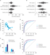
From Fig. 1A, the expression level of serum miR-221-3p in the gastric cancer group was significantly higher than the gastric polyp group and healthy control (P<0.01). The expression level in the gastric polyp group was significantly higher compared to the healthy control (P<0.01). miR-221-3p expression was significantly higher after operation than that before operation (P<0.01). Meanwhile, miR-122-5p expression in the gastric cancer group was significantly lower than the gastric polyp group and healthy control (P<0.01). The expression of miR-122-5p in the gastric polyp group was significantly lower than in the healthy control (P<0.01), and the expression after operation was significantly higher than that before operation (P<0.01), as shown in Fig. 1B.
Fig. 1
Clinical value of serum miR-221-3p and miR-122-5p levels in diagnosis and prognosis. (A, B) Serum miR-221-3p and miR-122-5p expression levels in each group. (A) Serum miR-221-3p expression levels in each group, (B) serum miR-221-3p expression levels in each group. Compared to healthy control, *P<0.01; compared to gastric polyp group, †P<0.01; compared to pre-operation, ‡P<0.01.(C) Correlation between serum miR-221-3p and miR-122-5p expression in gastric cancer. (D) Comparison of serum miR-221-3p and miR-122-5p expression in the area under the curve of diagnosis of gastric cancer. (E) Clinical prediction value of death within 2 years with serum miR-221-3p and miR-122-5p levels. Compared to survival group, *P<0.01. (F) Comparison of serum miR-221-3p and miR-122-5p levels in predicting death within 2 years.
Correlation between serum miR-221-3p and miR-122-5p expression in gastric cancer
As shown in Fig. 1C, miR-221-3p expression in gastric cancer was negatively correlated with miR-122-5p expression (r=−0.778, P<0.01).
Diagnosis efficacy of serum miR-221-3p and miR-122-5p expression in gastric cancer
From Table 1 and Fig. 1D on the diagnosis efficacy of gastric cancer, miR-221-3p diagnostic sensitivity was 71.6%, specificity was 82.7%, and the area under the curve (AUC) was 0.837. miR-122-5p diagnostic sensitivity was 91.5%, specificity was 73.6%, and the AUC was 0.881. The 2 makers of the binary regression equation are 0.041×XmiR-221-3p-0.084×XmiR-122-5p+2.884, the sensitivity of the combined detection was 91.5%, the specificity was 82.7%, and the AUC of combined detection was significantly better than miR-221-3p (z=5.052, P<0.001) and miR-122-5p (z=2.963, P=0.003). There was no statistical difference between the 2 indicators (z=1.309, P=0.191).
Table 1
Diagnosis efficacy of serum miR-221-3p and miR-122-5p expression in gastric cancer

Association between serum expression levels of miR-221-3p and miR-122-5p and clinical indicators in patients with gastric cancer
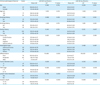
The serum miR-221-3p and miR-122-5p levels were not significantly associated with sex, age, smoking history, alcohol use history, family gastric cancer history, tumor location, pathological type, and tumor diameter (P>0.05, Table 2). Expression levels were significantly correlated with differentiation degree, TNM stage, lymph node metastasis, and invasion depth (P<0.01).
Table 2
Association between serum miR-221-3p and miR-122-5p expression levels and clinical indicators in patients with gastric cancer
Multivariate analysis of post-operative prognosis in patients with gastric cancer
All patients were followed up for more than 2 years until January 2019, with an average follow-up of 3.21±0.95 (2–5) years. The prognosis factors of sex, age, smoking history, alcohol use history, family gastric cancer history, tumor site, pathological type, tumor diameter, differentiation degree, TNM stage, lymph node metastasis, invasion depth, and miR-221-3p and miR-122-5p expression were analyzed using Kaplan-Meier analysis in patients with gastric cancer. The pathological type, tumor diameter, TNM stage, lymph node metastasis, invasion depth, and miR-221-3p and miR-122-5p expression were significantly correlated with 2-year survival rate (P<0.05). The significant variables with univariate analysis were included in the multivariate analysis using the Cox-Hazard model. miR-221-3p and miR-122-5p were independent prognostic factors for post-operative gastric cancer (P<0.01, Table 3).
Table 3
Multivariate prognosis analysis in gastric cancer patients

Association between serum miR-221-3p and miR-122-5p levels and follow-up after operation
After a 2 years follow-up, 22 patients died (death group) and 119 survived (survival group). From Fig. 1E, the serum miR-221-3p expression level in the death group was significantly higher than the survival group (P<0.01) and the serum miR-122-5p expression level was significantly lower than the survival group (P<0.01).
Clinical prediction value of death within 2 years with serum miR-221-3p and miR-122-5p levels
Data presented in Table 4 and Fig. 1F show that the sensitivity of serum miR-221-3p level in predicting death within 2 years in gastric cancer was 72.7%. The specificity was 87.4%, and the AUC was 0.838. The sensitivity of serum miR-122-5p was 72.7%, the specificity was 84.0%, and the AUC was 0.834. The equation of combined detection was 0.037×XmiR-221-3p-0.099×XmiR-122-5p-1.319 based on binary regression. The sensitivity of combined detection was 81.8%, the specificity was 94.1%, and the AUC was significantly higher than miR-221-3p (z=2.383, P=0.017). However, it was not significantly different from miR-122-5p (z=1.882, P=0.060). There was no significant difference between the 2 markers (z=0.066, P=0.946).
Table 4
Clinical prediction value of death within 2 years with serum miR-221-3p and miR-122-5p levels

Effect of miR-221-3p and miR-122-5p transfection on proliferation, migration, and apoptosis of gastric cancer cells
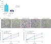
Gastric cancer cells showed significant expression of miR-221-3p and miR-122-5p after transfection of the miR-221-3p and miR-122-5p plasmids (Fig. 2A). SGC-7901 cell line showed the highest expression. Thus, the subsequent experiments were performed using the SGC-7901 cell line. As shown in Fig. 2B and C, the proliferation activity of gastric cancer cells transfected with miR-221-3p was significantly higher than that of the negative control at 48 hours and 72 hours (P<0.01), and the activity was significantly lower than the negative control after transfection with miR-221-3p inhibitor (P<0.01). The proliferative activity of gastric cancer cells after transfection with miR-122-5p was significantly lower than of the negative control at 48 hours and 72 hours (P<0.01). The proliferative activity was significantly higher than that of the negative control after transfection with miR-122-5p inhibitor (P<0.01) (Fig. 2B and D).
Fig. 2
Impact of gastric cancer cells on proliferation after transfection with miR-221-3p and miR-122-5p plasmids. (A) miR-221-3p and miR-122-5p expression in gastric cancer cells after plasmid transfection. Compared to MGC-803 and MKN28, *P<0.01. (B) Impact of miR-221-3p and miR-122-5p on proliferation of gastric cancer cells after transfection (72 hours×200). (C and D) Impact of miR-221-3p and miR-122-5p on proliferation of gastric cancer cells at different time points. (C) Impact of miR-221-3p on gastric cancer cell proliferation at different time points, (D) Impact of miR-122-5p on gastric cancer cell proliferation at different time points. Compared to 24 hours, *P<0.01.
There were significantly more gastric cancer cells in the lower chamber in the migration experiment than negative control after transfection of miR-221-3p (P<0.01, Fig. 3A and B). The number of gastric cancer cells decreased significantly compared to negative control after transfection with the miR-221-3p inhibitor (P<0.01). After transfecting miR-122-5p, as shown in Fig. 3A and C, the number of gastric cancer cells was significantly decreased compared to the negative control (P<0.01). After transfection of miR-122-5p inhibitor, the number of gastric cancer cells was significantly higher than of the negative control (P<0.01).
Fig. 3
Impact of gastric cancer cells on migration and apoptosis after miR-221-3p and miR-122-5p plasmid transfection. (A) Impact of miR-221-3p and miR-122-5p on migration of gastric cancer cells after transfection in upper chamber (24 hours×200). (B and C) Impact of transfection of miR-221-3p and miR-122-5p plasmids on gastric cancer cell migration for 24 hours. (B) miR-221-3p on migration of gastric cancer cells, (C) miR-122-5p on migration of gastric cancer cells. Compared to negative control, *P<0.01. (D) Gastric cancer cell apoptosis using flow cytometry after 24 hours of cell culture. (E and F) Apoptosis percentage of gastric cancer cells using flow cytometry after 24 hours of cell culture. (E) miR-221-3p on apoptosis percentage of gastric cancer cells, (F) miR-122-5p on apoptosis percentage of gastric cancer cells. Compared to negative control, *P<0.01.
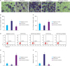
The apoptosis percentage in gastric cancer cells after transfection with miR-221-3p plasmid was significantly lower than that in the negative control (P<0.01), and the percentage was significantly higher than the negative control after transfection with miR-221-3p inhibitor (P<0.01, Fig. 3D and E). The apoptosis percentage of gastric cancer cells transfected with miR-122-5p plasmid was significantly higher than negative control (P<0.01), and it was significantly lower than negative control after transfection with miR-122-5p inhibitor (P<0.01) (Fig. 3D and F).
DISCUSSION
Increasing the early diagnosis rate of tumors is one method to improve the survival rate and prolong survival time. At present, the study of early diagnosis mainly focuses on the search for biomarkers in body fluids such as blood, chest fluid, and ascites. Relative to traditional tissue-based diagnostics, the body fluid sample is relatively easy to obtain and causes less harm to the patient. Since Mitchell et al. [18] confirmed that miR-141 in plasma can be used as a diagnostic marker for prostate cancer, new circulating miRNAs have emerged as tumor diagnostic markers. Serum levels of miR-18a-5p, miR-21-5p, miR-29a-5p, miR-92a-5p, miR-143-5p, and miR-378-5p were reported to be significantly reduced in patients with colorectal cancer [19]. Four plasma and 4 serum miRNAs were identified in the miR-106a-363 cluster, which is a novel biomarker in breast cancer diagnosis [20]. The combined detection of circulating miR-21 and let-7a has important diagnostic value in diagnosing non-small cell lung cancer [21]. miRNAs are stable in plasma, and there was no change in plasma expression level of miRNAs even when it was boiled at 100°C for 10 minutes, kept at room temperature for different time intervals (4, 12, and 24 hours), stored at −80°C, or repeatedly frozen and thawed at room temperature [22]. There is a current hypothesis that miRNAs in plasma are present in exosomes, complexes, or microvesicles secreted by tumor cells [23]. Some researchers believe that miRNAs are secreted by ceramide-dependent mechanisms that can be transferred to receptor cells [24], and they are increasingly used in understanding the mechanism, diagnosis, and prognosis of tumor cells.
In our study, the expression level of serum miR-221-3p in patients with gastric cancer was significantly higher than in the gastric polyp group and healthy control group, and the level in the gastric polyp group was significantly higher than in healthy control. The expression level of serum miR-221-3p increased significantly post-operation compared to pre-operation. This indicates that miR-221-3p is involved in gastric cancer development. This study also found that the serum miR-221-3p expression level was >105.26 fmol/L, the sensitivity was 71.6%, specificity was 82.7%, and AUC is 0.837 in the diagnosis of gastric cancer, indicating that miR-221-3p has a higher diagnostic efficiency. This study also found that serum miR-221-3p expression level was not associated with sex, age, smoking history, alcohol use history, family gastric cancer history, tumor location, pathological type, and tumor diameter. Serum miR-221-3p expression level was significantly correlated with differentiation, TNM staging, lymph node metastasis, and infiltration depth. It is indicated that serum miR-221-3p is involved in gastric cancer development, and it has a significant correlation with tumor staging. It was reported that exosomes transfer miR-221-3p from cancer cells to vascular endothelial cells and promote angiogenesis by down-regulating thrombospondin-2 [25] and transferring into human lymphatic endothelial cells to promote lymphangiogenesis and lymphatic metastasis by down-regulating angiostatin-1 [26]. This suggests that the exosome miR-221-3p derived from cervical squamous cell carcinoma may be a novel diagnostic biomarker and therapeutic target. However, there are few studies on gastric cancer. This study found that miR-221-3p promoted the proliferation and migration of gastric cancer cells transfected with miR-221-3p and inhibited apoptosis of the transfected gastric cancer cells, indicating that the miR-221-3p is involved in gastric cancer. The biological characteristics of gastric cancer cells are significantly changed. This study found that the serum miR-221-3p expression level in patients who died within 2 years of the follow-up was significantly higher than that in the survival group. The sensitivity was 72.7% and specificity was 87.4% when serum miR-221-3p was >161.32 fmol/L. The AUC was 0.838, indicating that these indicators have high diagnostic efficacy in predicting gastric cancer patient death within 2 years. Early intervention for patients with poor prognosis has important clinical significance for improving prognosis. In a colon cancer study, patients with higher miR-221-3p levels had lower survival rates. It is an important prognostic indicator [10] that can help doctors better predict prognosis and guide treatment decisions for colon cancer.
Serum miR-122-5p expression in patients with gastric cancer was significantly lower than that in gastric polyp group and healthy control. Post-operation expression was significantly higher than that pre-operation, and it was significantly correlated with differentiation degree, TNM stage, lymph node metastasis and invasion depth. This indicated that miR-122-5p was involved in gastric cancer development and associated with the degree of gastric cancer malignancy. Proliferation and migration of gastric cancer cells transfected with miR-122-5p was significantly inhibited whereas apoptosis was promoted in these cells; we obtained the opposite result after transfection with miR-122-5p inhibitor. This indicates that miR-122-5p is closely related to the proliferation, migration, and apoptosis of gastric cancer cells. Previous studies have shown that miR-122-5p is expressed in nasopharyngeal carcinoma cells. Cell proliferation, colony formation, cell migration, and cell invasion is inhibited by miR-122-5p, which targets specific nuclear matrix binding domain binding protein 1 [27]. In the study of cholangiocarcinoma, miR-122-5p was found to inhibit the proliferation, invasion, and growth of cholangiocarcinoma cells by targeting fructose diphosphate aldolase A [28]. miR-122-5p inhibits gastric cancer cell proliferation and induces apoptosis by targeting MYC. It is believed that miR-122-5p may be involved in gastric cancer progression [17], and it may be a new therapeutic target to treat the disease. The study found that the serum miR-122-5p expression level was significantly lower in the death patients within 2 years than that in the survival group, indicating that miR-122-5p is significantly correlated with gastric cancer prognosis. In another study, serum miR-122-5p expression level in patients with clear cell renal cell carcinoma is significantly lower than healthy control, associated with poor clinical pathology parameters [15], and is a novel non-invasive prognostic biomarker. This study showed that the expression level of miR-221-3p in gastric cancer was negatively correlated with the expression level of miR-122-5p, and the expression levels of miR-221-3p and miR-122-5p were independent prognostic factors for post-operative gastric cancer. In the diagnosis of gastric cancer, the sensitivity of the combined detection was 91.5%, the specificity was 82.7%, and the AUC was 0.936. These statistics indicated significantly higher diagnostic efficacy than the single indicators (miR-221-3p and miR-122-5p). At the same time, the sensitivity of the combined detection in the prediction of death within 2 years in patients with gastric cancer was 81.8%, the specificity was 94.1%, and the AUC was 0.919. The combined detection was better than the single indicator, indicating that it has important diagnostic efficacy in predicting death within 2 years. We conclude that there is a relationship between miR-221-3p and miR-122-5p. miR-221-3p is like an oncogene, miR-122-5p is like a tumor suppressor gene, and both have important clinical significance in the diagnosis and prognosis of gastric cancer.
In conclusions, miR-221-3p and miR-122-5p are involved in gastric cancer development, and they have important clinical value in gastric cancer diagnosis and prognosis.
 XML Download