

 PDF
PDF ePub
ePub Citation
 Print
Print


Introduction
Dyspeptic disorders such as gastroesophageal reflux, gastritis, peptic ulcer disease (PUD), and gastric cancer (GC) are major medical conditions.1 PUD is usually associated with a reduced health-related quality of life, whereas GC is the fourth most common cancer and the second leading cause of cancer-related deaths worldwide.2,3 Host factors such as genetics and nutrition, and environmental factors such as Helicobacter pylori infection may be involved in the development of these conditions.1,4
H. pylori infection has been shown to be a major risk factor for the development of PUD and GC.5,6 However, despite several investigations, it is still not completely understood why the majority of infected people (80%~90%) carry and spread the bacterium while they are asymptomatic, or why only a small percentage of infected people develop peptic ulcers, whereas others develop GC.6
Host immune responses against H. pylori can result in chronic inflammation in the gastric mucosa, which in turn leads to the development of pathological conditions including PUD and GC.5,6
Vascular endothelial growth factors (VEGFs) are glycoproteins secreted by tumor cells that are the most important factors in angiogenesis and tumor metastasis.7 The VEGF family includes VEGF-A to F and placental growth factor.7,8 Studies have shown that VEGF-A and B play a key role in blood vessel growth, whereas VEGF-C and D are important for the growth of lymphatic vessels.9,10 The role of VEGFs, particularly VEGF-A, C, and D in promoting angiogenesis and metastasis of many cancers including GC, has been previously discussed.11,12 Moreover, inflammatory cytokines such as interleukin (IL)-1, IL-6, and tumor necrosis factor-alpha (TNF-α) are generally responsible for the epigenetic alteration of gastric epithelial cells.13 These cytokines induce the mediators of angiogenesis, including VEGF and IL-8, which promote angiogenesis in cancer. These mediators also promote angiogenesis during chronic inflammation such as cardiovascular disease, rheumatoid arthritis, diabetic retinopathy, delayed-type hypersensitivity, and asthma.14 It has been shown that VEGF-A expression is up-regulated in response to H. pylori infection.15 Indeed, H. pylori activates the c-Jun N-terminal Kinases (JNK) signaling pathway, which leads to transactivation of the VEGF-A promoter. VEGFs promote angiogenesis, which is a pathophysiological mechanism that can result in inflammatory and ulcerative epithelial lesions and malignant tumor growth and metastasis.15
To understand the role of VEGFs in the pathogenesis of H. pylori-related gastric abnormalities, the mRNA expression levels of VEGF-A and C were determined in patients with peptic ulcers or GC, and compared with those with non-ulcer dyspepsia (NUD).
Materials and Methods
1. Patients and sampling
Patients with dyspepsia who underwent esophagogastroduodenoscopy at Imam Hospital or Tooba Outpatient Clinic (Mazandaran University of Medical Sciences, Sari, Iran) were enrolled in the study. All samples were collected between January 2012 and December 2013. The study was approved by the ethics committee of Mazandaran University of Medical Sciences. Clinical history, demographic data, and written informed consent forms were obtained from all study subjects. None of the subjects had a history of chronic inflammatory or autoimmune disorders or treatment with H. pylori eradication therapy, nor did they receive any non-steroidal anti-inflammatory drugs for 2 weeks prior to enrollment. Among patients with GC, none had undergone surgery, radiotherapy, or chemotherapy, or received any other medical intervention before sample donation.
Based on the endoscopic and histopathological assessments, the patient samples were divided into three groups: NUD, PUD, and GC. The histological grade of the gastric tumors was determined based on the state of differentiation. PUD was defined as a circumscribed mucosal break (>5 mm in diameter, with apparent depth) in the stomach or duodenum, covered with exudates. H. pylori infection was diagnosed by histopathological examination (including Giemsa staining) and a positive result on the rapid urease test performed on at least one additional biopsy sample. Patients were considered H. pylori positive if the results of one or both diagnostic methods were positive, and H. pylori negative if the results of both methods were negative. Patients in NUD group were then divided into two groups: H. pylori positive and H. pylori negative. Tissue samples were obtained from all patients during endoscopy and preserved in RNALater (Qiagen, Phoenix, AZ, USA).
2. RNA isolation and cDNA synthesis
Each tissue specimen was homogenized using mortar and pestle at room temperature. Total RNA was extracted from the dissected tissues using commercial RNA extraction kits (RNeasy Minikit; Qiagen), according to the manufacturer's instructions. The quantity and quality of the extracted RNA were assessed using a nanodrop spectrophotometer (Thermo Fisher Scientific Inc., Waltham, MA, USA) and agarose gel electrophoresis, respectively. RNA (1 µg) was reverse-transcribed into complementary DNA (cDNA) using the RevertAid™ First-Strand cDNA Synthesis Kit (Fermentas, Pittsburg, PA, USA) primed with random hexamers as per the manufacturer's instructions.
3. Quantitative reverse transcriptase-polymerase chain reaction (qRT-PCR)
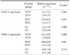
VEGF-A, VEGF-C and hypoxanthine-guanine phosphoribosyl transferase (HGPRT, for normalization), sequences were obtained from the GenBank (Table 1). Primers for amplification of VEGF-A, VEGF-C, and HGPRT were designed using the Beacon designer 7 software and synthesized by TIBmol (Germany) (Table 1).
qRT-PCR was performed using 96 well plates (Bio-Rad Laboratories Inc., Hercules, CA, USA) in a volume of 20 µl containing Maxima SYBR Green/ROX qPCR Master Mix (2×) (Thermo Scientific, Delaware, PH, USA), 10 pmol of each of the forward and reverse primers, and the appropriate amount of cDNA. The samples were denatured at 95℃ for 10 minutes, and then amplified during 40 cycles of: 95℃ for 30 seconds, 55℃ for 30 seconds, and 72℃ for 30 seconds on an iQ5 real-time thermal cycler (Bio-Rad Laboratories Inc.). Each sample was assayed in duplicate, and cycle threshold (Ct) values (corresponding to the number of PCR cycles at which the fluorescence emission monitored in real time exceeded a threshold limit [×10 the standard deviation of the baseline intensity]) were measured. A mean Ct value for each duplicate measurement was calculated. Relative gene expression was then calculated using 'ΔCt method using a reference gene' in the following manner for each sample: Ratio (reference/target)=2Ct (reference)-Ct (target).
4. Statistical analysis
Statistical analysis was performed using the SPSS Statistical Package ver. 17 (SPSS Inc., Chicago, IL, USA). The results were evaluated by using the independent sample t-test, the Mann-Whitney U test, and the Pearson and Spearman correlation tests where appropriate. Findings were considered significant when P-values were <0.05. The results presented in the text and tables represent the geometric mean in the case of 2-ΔΔCt, and the mean±standard error in the case of other variables.
Results
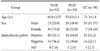
Fifty-two patients with NUD, 50 with PUD, and 38 with GC were enrolled in this study (Table 2). H. pylori infection was diagnosed in 29 (55.7%) NUD patients, 34 (68.0%) PUD patients, and 21 (55.2%) GC patients.
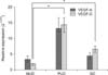
1. Relative expression of VEGF-C
Gene expression levels of VEGF-C were determined using qRT-PCR and normalized to the expression level of HGPRT for each individual sample. The results show that the relative expression levels of VEGF-C were higher in GC and PUD patients than in NUD patients (P<0.000 and P<0.000, respectively; Table 3, Fig. 1).
2. Relative expression of VEGF-A
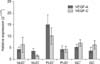
Gene expression levels of VEGF-A were also measured using qRT-PCR and normalized to the expression level of HGPRT for each individual sample. Patients with PUD or GC showed higher VEGF-A expression levels than patients with NUD, but the differences were not statistically significant (P=0.201 and P=0.217, respectively; Table 3, Fig. 1). Moreover, VEGF-A was expressed at higher levels in H. pylori positive patients than in H. pylori negative patients in all three groups; however, these differences did not reach statistical significance (P=0.164, P=0.927, and P=1.000, respectively; Table 4 and Fig. 2).
3. Correlation between VEGF-A and VEGF-C expression
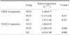
Positive correlation was found between VEGF-A and VEGF-C expression in patients with PUD (r=0.458, P<0.000) or GC (r=0.38, P<0.000), but not in patients with NUD (Table 5).
Discussion
This study evaluated the expression levels of VEGF-A and C in patients with PUD or GC compared with NUD patients as a control group. The findings showed increased expression levels of both VEGF-A and C in patients with PUD or GC compared with those in patients with NUD, although only the differences in the VEGF-C expression levels were statistically significant.
VEGF-C is a glycoprotein secreted by tumor cells and binds to receptors such as VEGFR-2 and VEGFR-3 that are found on the surface of endothelial cells in the lymphatic vessels. The binding of VEGF-C to its receptors leads to dimerization of these receptors, activation of their tyrosine kinase tails, activation of the serine protease and plasminogen activator, and ultimately the production of collagenase, which results in angiogenesis, particularly in the lymphatics.16,17,18
Previous studies have shown the expression of VEGF-C in tissues such as the placenta, ovary, small intestine, skeletal muscle, colon, and spleen.19,20 Karpanen et al.21 showed that due to its association with the growth of lymphatic vessels around the tumor, VEGF-C can promote growth of cancer cells and metastasis to the lymph nodes. Several studies have also demonstrated the overexpression of VEGF-C in metastatic tumors of the head and neck, thyroid, prostate, stomach, colorectal, and lung.20,22,23,24,25,26 Another study examined the importance of blood and lymphatic vessel growth factors, especially VEGF-C, in the growth and metastasis of tumor cells in patients with GC, and showed that increased expression of the VEGF-C glycoprotein was associated with increased tumor size and lymph node metastasis.18 Furthermore, using inducible mouse tumor models, silencing of the VEGF-C gene resulted in a significant reduction of tumor size in the experimental mice compared to that in the control mice.18 In addition, several studies have shown that VEGFs produced by the tumor cells can suppress the maturation of antigen-presenting cells, especially dendritic cells, which may lead to immune evasion and tumor progression.27 In our study, similar to previous studies on GC,12,20 we found significantly higher VEGF-C expression levels in patients with GC than in those with NUD.
We also found a significant increase in the VEGF-C expression levels in patients with PUD compared to those in patients with NUD. It has been shown that pro-inflammatory cytokines such as IL-1, IL-6, IL-8, and TNF-α can enhance the binding of nuclear factor-κB to the VEGF gene promoter leading to the increased expression of VEGF. This further increases the expression of intercellular adhesion molecule-1 and vascular cell adhesion molecule-1 on the surface of vascular endothelial cells, and the migration of immune cells such as B cells, T cells, NK cells, and macrophages, which results in increased inflammation.28,29,30 Consistent with the present study, another study evaluated the effects of the suppression of VEGF and angiopoietin expression in rats with peptic ulcers and found decreased production of pro-inflammatory cytokines, which resulted in reduced inflammation and wound severity, supporting the importance of VEGF and angiopoietin in the process of inflammation and ulceration.31
Because of the increased expression of VEGF-C in both GC and PUD patients, we assumed that expression of this glycoprotein is increased not only during cancer metastasis but also during chronic inflammation.29,32 Thus, the factors that cause inflammation, including infectious agents and carcinogens, may also lead to chronic inflammation by stimulating the production of proinflammatory cytokines and inducing production of VEGF-C. Therefore, the production of VEGF-C in peptic ulcers can lead to the progression of inflammation and development of cancer.
VEGF-A, on the other hand, is an inducible cytokine that promotes the growth of blood vessels and is a heparin-binding glycoprotein. The binding of VEGF-A to its specific receptors, including VEGF 1 and 2, results in the induction of mitosis and angiogenesis in vascular endothelial cells. Moreover, VEGF-A has an important role in metastasis occurring via the blood vessels.33
Similar to our study, George et al.33 showed that VEGF-A expression levels were increased in the sera from patients with colorectal cancer, suggesting that VEGF-A was involved in the progression of this malignancy. Another study also showed increased VEGF expression level in the advanced stages of GC compared to that in the earlier stages.34
In our study, despite the increased expression of VEGF-A observed, the differences between GC patients and controls were not statistically significant. Our findings could be influenced by the fact that we enrolled patients who had recently been diagnosed with GC, most of whom had early-stage disease with typical lower VEGF-A expression levels.
In addition, our results showed that the expression levels of both VEGF-A and VEGF-C were higher in H. pylori positive patients with PUD or GC than in H. pylori negative patients of the same groups, although this increase was also not statistically significant. One study showed that H. pylori induced the expression of VEGF-A via the phosphorylation of MEK/ERK transactivators and the activation of the JNK cascade.35 In that study, the binding of SP1 and SP3 proteins to the VEGF-A gene promoter stimulated VEGF-A expression. Furthermore, the same study showed that H. pylori strains with the cytotoxicity-associated gene (cag) pathogenicity island can activate the JNK cascade, while cagnegative strains cannot activate this pathway. Indeed, cag is a type IV secretion effector of H. pylori that is closely associated with the development of GC.35 These findings suggest an important role for cag-positive H. pylori strains in the production of angiogenic factors that lead to cancer metastasis.15 Although we did not examine the presence of cag in our samples, the prevalence of cag positive H. pylori strains is approximately 57% in patients infected with H. pylori, based on a 2012 study by Ajami et al.36 in the north of Iran.
In the present study, no significant increase in the VEGF-A and VEGF-C expression was found in the H. pylori positive patients compared with that in the H. pylori negative patients. This could be because our study did not differentiate between cag-positive and cag-negative strains of H. pylori. In addition, the unequal numbers of patients in the H. pylori positive and H. pylori negative groups might have biased the results.
The present study also determined the correlation between the expression levels of VEGF-A and VEGF-C by Pearson's correlation coefficient, which showed a significant positive correlation between the two variables. In agreement with our results, several studies have shown that VEGF-A and C have a synergistic effect, such that that production of one factor can stimulate the production of the other.37 Moreover, a study performed on patients with colorectal cancer33 showed a significant correlation between these two glycoproteins.
In summary, we report that inflammation of the gastric mucosa may result in the up-regulation of VEGF-C expression, which in turn, plays a role in the development of gastritis, peptic ulcers, premalignant changes, and ultimately, GC. The use of other techniques such as immunohistochemistry in addition to real-time PCR would provide a more accurate assessment of VEGF-A and VEGF-C protein levels, and likely demonstrate the increased expression of these glycoproteins. There were no follow-up studies on these patients, and thus we did not assess the expression of these glycoproteins during the various disease stages.


 XML Download