

 PDF
PDF ePub
ePub Citation
 Print
Print


Introduction
Gastric duplication cyst is a rare congenital anomaly of the gastrointestinal tract and is especially uncommon in adults.1 While most cases are diagnosed within the first year of life, a few cases have been diagnosed in adulthood.1,2,3,4 About one third of patients with gastric duplication cysts also have other congenital anomalies, such as annular or heterotopic pancreas, or vertebral anomalies such as spina bifida.5
Gastric duplication cysts might present with abdominal symptoms such as a palpable abdominal mass, pain, vomiting, and weight loss. In adults, most cases are discovered incidentally on radiological examination or gastric endoscopy.6
Accurate diagnosis of these cysts before resection is difficult. Differential diagnoses are varied, including gastrointestinal stromal tumors (GISTs), neuroendocrine tumors, pancreatic heterotopia, pancreatic pseudocysts, and neurogenic tumors. Malignant transformation of a gastric duplication cyst is very rare.7,8,9
We present three cases of asymptomatic noncommunicating gastric duplication cysts in adults and describe the endoscopic appearance as well as findings on computed tomography (CT) and pathological examination of these cysts.
Case Report
1. Case 1
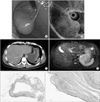
A 55-year-old man was admitted with a gastric submucosal tumor detected on esophagogastroduodenoscopy during a regular checkup without any abdominal complaint. Laboratory tests were normal. Esophagogastroduodenoscopy revealed a 7-cm protruding mass with a bridging fold at the cardia of the stomach (Fig. 1A). Endoscopic ultrasonography showed a homogeneous hypoechoic lesion located outside the gastric wall layers (Fig. 1B).
The patient underwent CT, which revealed a 7.9-cm well-marginated cystic lesion suspected to be a bronchogenic cyst or GIST (Fig. 1C). Magnetic resonance imaging (MRI), performed for accurate characterization, showed a 7.9-cm cystic lesion, with heterogeneous signal intensity on T2-weighted images, located in the submucosal layer of the posterior wall of the high body (Fig. 1D). Differential diagnoses including GIST, neuroendocrine tumor, and ectopic pancreas were made on the basis of the results of radiologic tests.
For treatment, the patient underwent gastric wedge resection, and a unilocular cystic mass measuring 7.0×7.0 cm was identified in the proper muscle layer. The resection margin was clear. Histopathological analysis of the resected tissue revealed a submucosal unilocular cyst measuring 6.0×6.0×4.0 cm, which led to the diagnosis of gastric duplication cyst (Fig. 1E, F).
2. Case 2
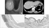
A 43-year-old woman presented with an asymptomatic gastric tumor detected during a medical checkup a year before admission. Because the size of the mass increased over the period of a year, we decided to perform a resection. There were no abnormal laboratory findings. Esophagogastroduodenoscopy revealed a 2.5-cm gastric tumor near the esophagogastric junction (Fig. 2A), and biopsy showed chronic gastritis. Abdominal CT showed an ovoid submucosal cystic tumor, which we suspected was a cystic hygroma or duplication cyst (Fig. 2B). Laparoscopic gastric wedge resection was performed. The mass protruded into the gastric lumen near the esophagogastric junction. There was no erosion or ulceration of the gastric mucosa near the mass. Histological examination of the resected mass revealed that the tumor was an intramural cyst consistent with a foregut duplication cyst lined by respiratory epithelium (Fig. 2C, D).
3. Case 3
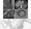
A 20-year-old woman presented with an asymptomatic gastric tumor detected during a medical checkup. She had no history of medical problems. Esophagogastroduodenoscopy revealed a protruding mass at the fundus of the stomach (Fig. 3A). CT showed a 4.8-cm dumbbell-shaped mass at the gastric fundus, consistent with either a submucosal tumor or a neurogenic tumor (Fig. 3B). The tumor had inner cystic components, but the adjacent lymph nodes were not enlarged. Therefore, there was a higher probability of the tumor being a neurogenic tumor than a GIST. Laparoscopic gastric wedge resection was performed (Fig. 3C). The resected mass was dumbbell shaped and filled with a mucus-like yellowish fluid (Fig. 3D). Pathological analysis showed that the cyst was lined by gastric foveolar epithelium with pyloric glands and two or three complete layers of smooth muscle bundles, which suggested a duplication cyst (Fig. 3E, F). There were four reactive lymph nodes. More than two years have passed since the resection, and no apparent problems have been detected with gastroscopy so far.
Discussion
The first case report of a gastric duplication cyst was published in 1911 by Wendel.10 Gastric duplication cysts are spherical hollow structures with a smooth muscle coat, lined by the mucous membrane.11 They are rare congenital malformations of the alimentary tract and most commonly occur in the ileum, followed by the esophagus, jejunum, colon, stomach, and appendix.1,2,4
Most gastric duplication cysts are located along the greater curvature of the stomach, adjacent to the gastric wall, because these duplications are usually found to be distributed dorsally to the primitive gut during development.4 As observed in the previous studies, all three cases in the present study were located along the greater curvature, near the esophagogastric junction of the stomach.
The etiology of gastric duplication cysts remains unclear. Histologically, these cysts have an inner lining of gastric mucosa and an outer smooth muscle coat. These malformations are believed to be formed before differentiation of the lining epithelium, and the respiratory epithelium is the inner lining epithelium in some gastric duplication cysts, as observed in the second case in the present study.12 In this case, the term 'foregut duplication' is preferred, because the term 'gastric duplication' denotes the presence of the gastrointestinal mucosa. The second case in this study, for example, was a foregut duplication cyst and not a gastric duplication.
Gastric duplication cysts might present with abdominal symptoms including a palpable abdominal mass, pain, vomiting, and weight loss. These cysts are usually asymptomatic in adults, and consequently, they are diagnosed incidentally, as observed in the cases in this study. However, in some cases, symptoms such as massive bleeding from the cysts13 have been observed, and malignant transformation of these cysts, while rare, has been reported.8 Therefore, resection might be the first treatment option. Gastric duplication cysts can be removed using minimal invasive laparoscopic techniques or an open method; however, several physicians have also reported the use of endoscopic treatment.14,15,16 In the present study, we believed that minimally invasive surgery would not be beneficial because of the large size of the tumors. Aspiration of the cyst fluid might be helpful in endoscopic or laparoscopic procedures.
In this study, although the first patient underwent esophagogastroduodenoscopy, CT, and MRI, we had to exclude the possibility of a GIST. CT findings in the third case showed features similar to those of a GIST or neurogenic tumor. Various differential diagnoses were suggested. Endoscopic and endoscopic ultrasound findings of small subepithelial lesions suggested the potential diagnosis of GIST, neuroendocrine tumor, or pancreatic heterotopia. Although fine needle aspiration cytology might be useful for diagnosis,17,18 there is no accurate diagnostic tool for preoperative diagnosis. Moreover, aspiration-associated complications such as fistula formation and hemorrhage have been reported.19
If the possibility of a solid tumor such as a GIST or neurogenic tumor is excluded before operation, intraoperative cyst aspiration can be attempted to decrease the size of the tumor so that laparoscopic surgery can be performed. Although there are no accurate diagnostic tools for such cases, surgical resection and pathological confirmation can be effective to not only make a definite diagnosis but also guard against the unknown risk of malignant transformation.
In conclusion, a gastric duplication cyst is a rare congenital anomaly that is difficult to diagnose definitively. It should be considered as a differential diagnosis of gastric cystic lesions.
 XML Download