

 PDF
PDF ePub
ePub Citation
 Print
Print


INTRODUCTION
A cecal volvulus is caused by abnormal mobility of the cecum, resulting from improper developmental fusion of the cecum and ascending colon mesentery with the retro-peritoneum. Cecal volvulus is uncommon in pediatric patients, accounting for <1% of all cases of intestinal obstruction [1]. However, intestinal volvulus is a common cause of obstruction in cerebral palsy patients [2-4]. Few reports have described cecal volvulus with chronic constipation in lissencephaly. Here, we report the case of a patient with a history of mental disability presenting with bowel obstruction diagnosed as cecal volvulus.
CASE REPORT
A 19-year-old male (height, 145 cm; weight, 34 kg), who was treated for cerebral palsy and refractory epilepsy due to lissencephaly, visited our hospital (Yeungnam University Medical Center, Daegu, Korea) because of severe abdominal distension and vomiting. Also, this patient was not able to perform independent daily living without any assistance because of mental disability. The patient had been treated with an anti-epileptic agent such as lamotrigine, phenobarbital, leviteracetam, rivotril, and ativan for epilepsy since he was a neonate and had visited our hospital several times for rectal decompression because of chronic constipation. The patient, suffered from chronic constipation over 3 years, had history of ≤2 bowel movements per week with hard large stool, necessitating rectal stimulation for defecation. Two months prior to admission, the patient had experienced a 3.0 kg weight loss and sleep disturbance with irritability.
Upon admission, the patient was lethargic with dried lips. Abdominal examination showed epigastric and lower abdominal distension with decreased bowel sounds, which had mild rigidity. We could not check the site of pain and tenderness due to impaired communication with the patient. Laboratory findings showed leukocytosis with a white blood cell count of 45,690/µL, increased C-reactive protein at 15.14 mg/dL (reference range <0.5 mg/dL), and increased blood urea nitrogen/creatinine at 33.15/1.91 mg/dL. The patient had an electrolyte imbalance consisting of hyponatremia (124 mmol/L Na+) and hyperkalemia (5.9 mmol/L K+). Intravenous antibiotics of 3rd cephalosporin (50 mg/kg/day) and metronidazole (15 mg/kg/day) were administrated.
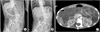
An initial abdominal x-ray showed a markedly distended stomach and a huge quantity of gas in right lower abdomen (Fig. 1A). We performed continuous suction with a naso-gastric and rectal tube for decompression and observed the patient. The next day, abdominal distension progressively deteriorated and a follow-up laboratory examination revealed uncontrolled inflammation. A follow-up supine abdominal x-ray 24 hours after the decompression revealed fixed haustral gas formation despite decompression with gastric and rectal tube insertion (Fig. 1B). Leukocytosis was improved, but C-reactive protein levels increased markedly to 34.11 mg/dL. An abdominal computed tomography scan without contrast enhancement scan showed dilated small bowel in the abdomen but no 'while' or 'bird peak' sign (Fig. 1C).
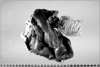
We suspected intestinal obstruction as the primary cause of the symptoms and planned an explorative laparotomy. During the laparotomy, ischemic changes were seen throughout the cecum and part of the ascending colon. The cecum, which was not fixed at the retro-peritoneum, was dilated and twisted around the terminal ileum (Fig. 2) and an approximately 20 cm length segmentectomy of the proximal ascending colon was completed with preserved ileocecal valve.
The patient was admitted to the intensive care unit and hospital ward for >2 months due to post-operative ileus. Approximately 3 months after the laparotomy, previous ileus signs had nearly disappeared and the patient's symptoms were much improved.
DISCUSSION
A cecal volvulus is defined as the rotation or torsion of a flexible cecum and ascending colon, frequently progressing to bowel obstruction, ischemia, necrosis, and perforation [5]. Volvulus of the cecum is uncommon in adults and extremely rare in childhood. It accounts for <1% of all cases of intestinal obstruction and 25-40% of the cases of volvulus of the colon [6]. A variety of factors affect colonic volvulus, including bowel distention, adhesions from previous surgery, mal-rotation, pregnancy, weight loss, and chronic constipation [7]. Cecal volvulus in children with mental disability is associated with aerophagia and constipation, which induces bowel distention. Severe stool accumulation in the large bowel of mentally impaired children is common due to insufficiently treated chronic constipation, which results in a heavy and extremely dilated bowel [8]. The patient in this case had developmental delay and chronic constipation with a history of several rectal decompressions. Sequentially, mental disability after lissencephaly might result in the chronic constipation, which and recent weight loss affects the cecal volvulus. Also, as the cause of the chronic constipation, anti-epileptic drug as well as mental disability may be considered.
Lissencephaly or agyria is a rare anomaly characterized by incomplete or failed neuronal migration during gestational weeks 12-24 that causes lack of gyri and sulci development, as well as developmental delay and cerebral palsy [9]. Impaired communication and behavior, motor and sensory impairments and altered reaction to pain can result in a late diagnosis of lissencephaly susceptible patients. Children with colonic volvulus typically present with an acute onset of abdominal pain and obstruction or chronic intermittent symptoms such cramps, bloating, nausea and vomiting that increase in severity [10]. These symptoms occur quite frequently in children with neuro-developmental delay, and intestinal obstruction usually responds to an enema and laxatives. Those methods, however, are not effective in severe colonic volvulus [10].
In 19-50% of cecal volvulus cases, cecal resection is performed as a consequence of cecal gangrene and/or perforation. Non-surgical management (e.g., colonoscopy, reduction barium enema) for de-torsion of a cecal volvulus is not recommended, as these treatments result in a 20-25% risk of concurrent cecal necrosis, have failure rates approaching 95%, and have an increased risk of colon perforation [11]. A high index of clinical suspicion with early diagnosis and intensive peri-operative care is recommended to reduce high morbidity and mortality in cases of bowel obstruction in cerebral palsy patients [6].
In the patient with a history of neuro-developmental delay such as lissencephaly who was followed by recurrent constipations, the patient should be considered that rare cecal and proximal large bowel volvulus might be the cause of progressive abdominal distension, although typical abdominal pain and terderness cannot be checked for the mental disability.
 XML Download