

 PDF
PDF ePub
ePub Citation
 Print
Print


Introduction
The vas deferens (VD) is the distal continuation of the epididymis. It starts as a muscular tube at the epididymal tail. Approximately 45 cm long, the VD conveys sperm to the ejaculatory ducts. Initially it runs along the posterior aspect of the testis medial to the epididymis. From the upper pole of the testis it ascends into the posterior part of the spermatic cord and traverses the inguinal canal to enter the pelvis. It runs retroperitoneally in the pelvis and finally joins with the duct of the seminal vesicle to form the ejaculatory duct [1].
Duplication of VD is a rare congenital anomaly [2-7]. A recent review on this anomaly identified a total of 22 cases of VD duplications, most of which were identified incidentally [8]. Duplication of VD was most commonly identified during procedures such as orchiopexy, inguinal hernia repair, vasectomy, varicocelectomy, and radical prostatectomy [2-8]. Duplication of the VD may be associated with unilateral renal agenesis and other renal anomalies [8]. Recognition and interpretation of this anomaly is important to avoid injury to the VD or an ineffective vasectomy. We report a case of duplicated VD noted in a formalin embalmed cadaver, and discuss its embryological perspective and clinical importance.
Case Report
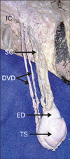
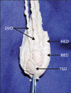
We discovered a rare congenital anomaly of unilateral duplication of the VD in the right spermatic cord of an adult cadaver of South Indian origin during regular dissection classes for first year medical students. Careful dissection of the right spermatic cord revealed 2 separate and completely developed cord like structures (Fig. 1). One of these cords was smaller and showed a thinner muscular wall. When traced caudally, the cords were found to be communicating separately with the tail of the epididymis (Fig. 2). Cranially, both traversed the inguinal canal as content of the spermatic cord, and finally fused at the level of the deep inguinal ring. Subsequent dissection of the scrotum revealed a normally located testis and epididymis (Fig. 2). The left spermatic cord and VD were completely normal. No associated renal abnormalities were noted.
Discussion
Instances of VD anomalies are very rare, estimated to occur in less than 0.05% of the general population [3]. Reported VD anomalies include complete absence, duplication, ectopia, hypoplasia, and diverticulum [9]. Duplication of VD is a rare congenital anomaly, with a total of 22 cases described so far in medical literature [8]. This anomaly may be associated with other congenital abnormalities such as ipsilateral renal agenesis and cystic fibrosis, but no cases of abnormalities with the kidneys, ureters, or bladder have been reported [8]. Duplication of the VD and double VD are 2 different terms. According to Vohra and Morgentaler [9], duplication of the VD is defined as a second VD within the spermatic cord whereas double VD describes an ectopic ureter draining into the ejaculatory system [9]. Among the reported cases where bilateral spermatic cord examination was done, there was an equal distribution of right-sided, left-sided, and bilateral duplication of the VD [8]. Poly-vasa deferentia is classified into 3 types. In type I, a second VD is identified in the spermatic cord with no polyorchidism. Type II multiple VD is associated with polyorchidism. Type III represents double VD, where an ectopic ureter drains into the ejaculatory system [8]. The present case, with a second VD in the spermatic cord and no polyorchidism, belongs to type I.
Duplication of VD is said to be complete when the 2 VDs are totally separated from the tail of epididymis to the prostate. If they are separated for only a short distance, then the duplication is said to be partial. In all previously reported cases, partial duplication occurred in 53% of the cases, and duplication was complete in 47% [8]. The etiology of these variations can be explained by understanding the embryological derivation of the VD. Development of the male reproductive organs begins at approximately the 7th week of gestation. At this stage the paramesonephric duct regresses and the spermatogenic cord appears in the fetal testis [9]. The main reproductive ducts such as the epididymal canal, VD, and ejaculatory duct are derived from the mesonephric duct, and the central portion of the mesonephric duct gives rise to the VD. Duplication of the VD may result from duplication of the fetal mesonephric system [4, 9] or transversal division of the mesonephric duct during organogenesis [2].
All reported medical cases of duplication of the VD were identified incidentally during clinical evaluation [8]. Mysorekar [10] identified this anomaly in a cadaver during autopsy. He noted that one of the duplicated VD was smaller and had a narrow lumen and a thinner muscle coat [10]. In the present case, we found a duplicated VD where 2 separate ducts arose separately from the tail of the epididymis and fused at the level of the deep inguinal ring, after traversing the inguinal canal. We also observed that 1 of the duplicated VD was smaller and had a thinner muscular coat.
It is important for the surgeon to be familiar with this anomaly in order to prevent iatrogenic injury to the VD during repair of the inguinal hernial sac and avoid inadvertent failure of a vasectomy procedure [8]. Precise dissection of inguinal canal contents during varicocele surgery is extremely important, as failure to recognize a duplicated VD in the inguinal canal may cause inadvertent damage to abnormally located, but functionally important, structures [3].
 XML Download