

 PDF
PDF ePub
ePub Citation
 Print
Print


Introduction
The digastric muscle, as the landmark in head and neck surgery, has two bellies with separate embryological origins and innervations. The posterior belly takes its origin from the medial side of the mastoid process running down and forward to the hyoid bone. The anterior belly originates from the inferior border of the mandible, just close to the midline, and runs backward superficial to the mylohyoid muscle. These two bellies are united by an intermediate rounded tendon, which attaches to the body and greater horn of the hyoid bone.
As variations in the digastric muscle are very common, they have been studied and classified by many authors [1-4]. According to Kim et al. [5], accessory bellies of the anterior belly of the digastric muscle occur in 23.5% of Koreans; however, unique cases have been reported in recent years [6-8]. Surgical advances in this region have led to an increased demand for more information concerning muscle variations that may provide precise anatomic data for therapeutic approaches [5, 8]. Therefore, it is necessary to recognize anatomic variations of the digastric muscle in order to avoid misdiagnoses and complications during therapeutic procedures. In this report, we describe a unique variation of bilateral digastric muscle and discuss possible surgical complications.
Case Report
During the dissection, we found bilateral variations of the digastric muscle in the cadaver of a 74-year-old Korean male. We carefully removed the skin, subcutaneous tissue, fascia superficialis, and platysma, exposing the muscle in the submental region. The muscle in the submental region was made apparent. Asymmetric variation of the digastric muscle was found, and origins, insertions, shapes, blood supplies, and innervations were also investigated.
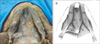
Exposure of the digastric triangle revealed four anterior bellies, two accessory medial bellies and two main anterior bellies (Fig. 1). Both accessory bellies originated from the medial side of normal anterior bellies posterior and medially. Then, it merged and attached at the mylohyoid raphe of the mylohyoid muscle. The right and left accessory bellies were approximately 30 and 32 mm in length, respectively, and both were 7 mm in width. The anterior and accessory bellies on both sides were innervated by the mylohyoid nerve on each side. Considering their courses and innervations, these accessory bellies were defined as accessory anterior bellies.
The 3rd accessory belly originated from the right intermediate tendon and continued for 15 mm further horizontally. It merged the right lower bundle of the right accessory belly and inserted at the mylohyoid raphe of the mylohyoid muscle together. It was innervated by the mylohyoid nerve and had no connection with the left diagstric muscle. The normal anterior and posterior bellies of the digastric muscles on both sides had normal origins, courses, and innervations. The submental artery supplied the muscles on each side.
Discussion
The digastric muscle stabilizes and regulates the position of the hyoid bone and assists jaw movements. It normally depresses the mandible and can elevate the hyoid bone. The imbalance between the muscles of the right and left sides may affect the movements of the hyoid bone and jaw [4]. Due to the clinical importance mentioned above, many authors have studied and classified variations in this muscle [1-5]. According to these studies, an additional anterior belly originated from the anterior belly itself, the intermediate tendon, the hyoid bone, the mandible, and the digastric fossa. Moreover, the insertion points included the mylohyoid raphe and even the mylohyoid muscle. Bilateral variations of the digastric muscles have been reported to be rarer than unilateral variations [3, 6, 9]. Therefore, bilateral asymmetric variation in this muscle as in the present case is rare and has clinical importance due to its effect on the movements of this region.
Previous studies were concerned about the number of supernumerary heads of the digastric muscle [1-3]. However, a recent study focused on the morphological types of this muscle based on their functional importance, and it was classified into digastric fossa type and crossover type depending on whether it crosses the median line or not [9]. They reported that digastric fossa type is more common and is usually bilateral. In digastric fossa type, one or two aberrant bundles are frequently found, and a case with bilateral four aberrant bundles was identified. In crossover type, bilateral variation is absent, and only unilateral aberrant bundles (1-3 bellies) are infrequently present.
Recently, unique bilateral cases have been reported by many authors [6, 9, 10]. Aktekin et al. [10] reported a bilateral symmetric variation with four accessory bellies gathered in a common tendon on the midline. In the present case, accessory bellies from the mandible medial to the origin of the anterior belly on both sides merged and attached at the mylohyoid raphe symmetrically. In addition, the 3rd accessory belly originated from the right intermediate tendon and merged at the right low margin of the right accessory belly. All of the accessory bellies coming either from the mandible or the intermediate tendon gathered in a common tendon on the midline, as in the case reported by Aktekin et al. [10]. However, an asymmetrical and unilateral belly connected to these two accessory bundles like our 3rd belly has not been reported in any study. Though Aktekin et al. [10] found a bilateral symmetric variation, they emphasized the clinical importance of bilateral asymmetric variation as in our case.
This presentation has anatomical significance due to its novelty and will guide clinicians during surgical procedures and radiological diagnoses. Variations in the anterior bellies of the digastric muscle can be easily confused with a mass or lymph node in computed tomography and magnetic resonance imaging. Since this connection may affect the movement of the mandible and hyoid bone, asymmetrical muscular variation could cause an inexperienced clinician to misinterpret pathological conditions. As a landmark during head and neck surgery, knowledge on variations of the anterior belly of the digastric muscle is very important for clinicians to avoid iatrogenic injuries or complications.
 XML Download