

 PDF
PDF ePub
ePub Citation
 Print
Print


Introduction
Pharyngoesophageal diverticuli are relatively rare diseases [123]: the Zenker's diverticulum is an outpouching from the muscular gap in the posterior portion above the cricopharyngeus muscle with an estimated incidence of less than 0.5% and the Killian-Jamieson diverticulum is an outpouching from a muscular gap in the anterolateral wall of the proximal cervical esophagus just below the cricopharyngeus muscle and superolateral to the longitudinal muscle of the esophagus with an incidence ratio of 1:4 as compared to Zenker's. Although simultaneously occurring Zenker's and Killian-Jamieson diverticula in one patient was reported [4], we showed a direct evidence of Killian-Jamieson diverticulum lined with two different epithelial cells in a Korean male cadaver.
Case Report
During a routine educational dissection at Jeju National University Medical School in 2017, we found a well-defined lateral diverticulum of the proximal esophagus in a 55-year-old Korean male cadaver, whose cause of death was “unknown.”
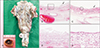
At the neck, superior, middle and inferior pharyngeal constrictor muscles were dissected. When the outer longitudinal layer of the esophagus was removed, a lateral outpouching (Fig. 1A) was detected on the lateral part just inferior to the transverse fibers of the cricopharyngeus muscle, the most inferior part of the inferior pharyngeal constrictor muscle. The lateral outpouching of a hollow structure (rectangle in Fig. 1A) had a dimension of 1.8×1.4×1.0 cm, and the thickness of its wall was about 2 mm.
Hitopathologically, the diverticular wall consisted of mucosa, muscle layer, and adventitia (Fig. 1B, C). The mucosal surface was lined by two types of epithelial cells: stratified squamous epithelium (Fig. 1D) and simple cuboidal or low-columnar epithelium (Fig. 1E). Multiple foci of superficial ulceration were observed. The muscular fiber was attenuated and haphazardly arranged. No epithelial dysplasia or malignant transformation was identified.
Discussion
Killian-Jamieson diverticulum is a thin-walled diverticulum, mean diameter 1.5 cm (range, 0.5–10 cm), which is usually considered as a false diverticulum without a muscular layer [35]. In this report, a typical Killian-Jamieson diverticulum was observed between the fibers of the cricopharyngeus muscle superiorly and longitudinal muscle of the esophagus inferiorly (so called Killian-Jamieson area [1]) with a diameter of 1.8×1.4×1.0 cm and haphazardly arranged and attenuated muscle fibers. Our case, however, had different property on the epithelial lining, stratified squamous epithelium and cuboidal to low-columnar epithelium, which has never been reported to the best of our knowledge.
There might be a possibility of the unrecognized or erroneously classified Killian-Jamieson diverticulum as Zenker's, because the majority of articles about hypopharyngeal diverticula have concentrated on Zenker's diverticulum. The lateral Killian-Jamieson diverticula, however, might be relatively common, with a range of 35 % [6] up to 44% [7] of hypopharyngeal diverticula. The incidence of the Killian-Jamieson diverticulum was reported 1.87% (16 of 854) [7] to 3.4% (17 of 500) [8] from dysphagia patients, which is a very similar incidence of Zenker's (2.34%, 20 of 854) [7].
Although the incidence of an occult malignancy (squamous cell carcinoma) in the wall of a long-standing Zenker's diverticulum has been reported to be 0.4% [9], the incidence of malignant transformation on Killian-Jamieson diverticulum has never been reported. Zenker's and Killian-Jamieson diverticula have similar histopathologic characteristics: therefore they might share common risk factors for malignancy, such as larger diverticula of long duration [9]. In addition, the occurrence of metaplasia to simple cuboidal to low columnar epithelium suggests that there might be a possibility of persistent stimulations developing into a dysplastic or malignant transformation. In this case, we carefully excluded the possibility of adenocarcinoma according to the literature that columnar lining esophagus without intestinal metaplasia is less associated with adenocarcinoma [10].
Taken together, awareness of the fact that Killian-Jamieson diverticula can be misdiagnosed as thyroid nodules [5] is important to avoid unnecessary interventions. The differentiation between Zenker's and Killian-Jamieson diverticula is also important, as surgical management differs [4]. In addition, physicians should aware the possibility of carcinogenesis in the Killian-Jamieson diverticulum, although no epithelial dysplasia or malignant transformation was identified in this direct evidence.
 XML Download