

 PDF
PDF ePub
ePub Citation
 Print
Print


TO THE EDITOR: Immunoglobulin M (IgM)-type monoclonal paraproteinemia is reportedly present in patients with various subtypes of lymphoma, but approximately 60% and 20% of cases are found in patients with lymphoplasmacytic lymphoma (LPL)/Waldenström's macroglobulinemia (WM) and chronic lymphocytic leukemia/small lymphocytic lymphoma (CLL/SLL), respectively [123]. Other types of non-Hodgkin lymphoma with serum monoclonal IgM paraprotein occur rarely, and we report here a case of nodal marginal zone lymphoma (NMZL) with diffuse bone marrow (BM) involvement and IgM-type monoclonal paraproteinemia.
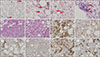
A 60-year-old woman visited our institution in December 2018 with the symptoms of fever, cough, and epigastric pain lasting for one week. Computed tomography (CT) showed the presence of mild hepatosplenomegaly without definite evidence of a splenic mass and multiple lymphadenopathies involving the bilateral cervical, axillary, and upper mediastinum lymph nodes (LNs). Whole-body positron emission tomography-CT scan results showed diffusely increased metabolism with marrow expansion and multiple hypermetabolic LNs in the bilateral cervical, axillary, mediastinal, and abdominal areas, but there was no definite evidence of increased metabolism in other extranodal lesions, such as the stomach or other intestines. Her hemogram results at the first visit were as follows: white blood cells, 14.6×10⁹/L; hemoglobin, 9.8 g/dL; and platelets, 73×109/L. The peripheral blood smear (PBS) revealed the presence of medium-sized to large neoplastic lymphoid cells exhibiting the absence of villous projection in their cytoplasm at a frequency of 32% (Fig. 1A).
The BM aspiration showed normocellular marrow with increased infiltration of large neoplastic lymphoid cells (8.5%) (Fig. 1B–D) and plasma cells (4.5%), and BM biopsy showed normocellular marrow (cellularity 60%) with diffuse infiltration of large neoplastic lymphoid cells (Fig. 1E, F). Subsequently performed immunohistochemical (IHC) staining of BM biopsy sections showed the presence of large neoplastic lymphoid cells with strong membranous positivity for cluster of differentiation (CD)20 (Fig. 1G), negativity for both CD3 and CD10 (Fig. 1H, I), but nuclear positivity for both B-cell lymphoma (BCL)-6 and multiple myeloma oncogene 1 (Fig. 1J, K). In addition, increased plasma cells showed positivity for CD138 (Fig. 1L). Interestingly, serum electrophoresis/immunofixation results showed the presence of monoclonal gammopathy, IgM kappa type with M-protein of 5.6 g/L.
Excisional biopsy specimens obtained from the left axillary LN showed an increase in reactive T cells with positivity for CD3, CD4, and CD8 on IHC staining and the presence of small neoplastic lymphoid cells with transformed large cells exhibiting positive results on CD20, BCL-2, and Ki-67 (40%) IHC staining, but negative results on CD10, Cyclin D1, and Epstein–Barr virus in situ hybridization IHC staining. Based on these results, the pathologic diagnosis of high-grade NMZL was made, and she was planned to receive intravenous methylprednisolone at 1 mg/kg for every 12 hours at first, followed by rituximab and bendamustine-based chemotherapy.
No studies have suggested the possible mechanism or hypothesis of IgM-type monoclonal paraproteinemia in B-cell lymphoma. However, in the association between the development of B cells and specific lymphoma subtypes, previous studies have demonstrated the association between monoclonal paraproteinemia and diffuse large B-cell lymphoma (DLBCL) with non-germinal center B cell (GCB) subtype with an explanation summarizing that DLBCL with non-GCB subtype develops from post-GCB and is associated with an up-regulation in Blimp-1, which is a master regulator of plasma cell differentiation [45].
A previous study showed that in 430 patients with serum IgM-type monoclonal paraproteinemia, 56.3% of the cases were of monoclonal gammopathy of undetermined significance (MGUS) and 16.5% were of WM, followed by other lymphomas, CLL, and primary amyloidosis with frequencies of 6.5%, 4.9%, and 1.4%, respectively [1]. Another previous study analyzed 106 patients with lymphoma and IgM-type monoclonal paraproteinemia and showed that although LPL/WM were the most common disease subtypes, 24 patients showed other lymphoma subtypes such as 10 patients with CLL, 5 patients with DLBCL, and 3 patients with extranodal MZL of mucosa-associated lymphoid tissue (MALT) lymphoma, follicular lymphoma (FL), and mantle cell lymphoma (MCL), respectively [2]. Another study analyzed 382 patients with IgM-type monoclonal paraproteinemia and lymphoma and revealed the diagnoses of LPL/WM, CLL/SLL, FL, MALT lymphoma, splenic MZL, MCL, and DLBCL with frequencies of 58.9%, 20.2%, 4.7%, 3.9%, 2.9%, 2.9%, and 1.8%, respectively [3]. This study demonstrated only one case of NMZL with IgM-type monoclonal paraproteinemia, representing a frequency of 0.003% [3], and to date, no cases have been reported except for a case study performed in China that presented two cases of NMZL with IgM-type monoclonal paraproteinemia [6]. Therefore, it can be speculated that although some cases of MZL with IgM-type monoclonal paraproteinemia have been reported, most cases are of the extranodal and splenic subtypes, and cases of NMZL are extremely rare. The mechanism or hypothesis that explains the rarity of IgM-type monoclonal paraproteinemia in NMZL and possibility in MZL of extranodal and splenic subtypes has not been suggested in a previous study, and this issue should be further analyzed in a more comprehensive future study. In summary, we report here a rare case of NMZL with diffuse BM involvement and IgM-type monoclonal paraproteinemia.
 XML Download