

 PDF
PDF ePub
ePub Citation
 Print
Print


Dear Editor:
Female pattern hair loss (FPHL) is a disease with genetic predisposition that is characterized by miniaturization of hair follicles followed by hair volume reduction. Contrary to male androgenetic alopecia, FPHL is less obviously related to androgen metabolism, moreover, the increased number of FPHL cases in women above age 50 may imply estrogen's stimulatory role in hair growth1. Several intercellular cascades were observed as dysregulated in FPHL, including transforming growth factor-β1 and stem cell factor/cKIT signalling23.
One of the most significant gene expression regulators at the posttranscriptional level are microRNAs which are capable of mRNA degradation and translation inhibition. Previous studies pointed out the role of microRNA in hair follicles normal functioning and pathological states' formation. Hence, miR-214 controls hair follicle morphogenesis by β-catenin targeting4. As for miR-203, it regulates the transcription factor p63 expression which is essential for hair follicle development5. Epigenetic alterations in FPHL are not clear yet but supposed to play an evident role.
Following the above-mentioned reports, we carried out the microRNA profiling in normal female scalp, normal male interscapular skin, and FPHL skin in order to identify differences in microRNA expression pattern and better understand FPHL pathogenesis as well as to define novel FPHL therapeutic targets.
The affected scalp skin of women with FPHL (n=3), at points 3~4 on the Sinclair scale aged 43~55 years, was obtained from equal frontal zone. Skin biopsies of control females (n=5) were taken from the frontal region of the scalp of healthy donors. Normal male skin tissue (n=5) was obtained from thoracic interscapular area of healthy voluntaries (IRB protocol No 70/2016 issued on June 6, 2016, demograhics and clinical data presented in Supplementary Table 1, 2). MiRNA expression profiles were generated by using Affymetrix GeneChip® miRNA microarray. Full experimental details are provided in the Supporting Information.
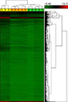
Microarray analysis revealed 981 up-regulated microRNAs at FPHL versus female controls and 972 microRNAs as up-regulated as compared to those of normal male skin whereas none of microRNAs were down-regulated. MiR-197-3p, miR-3613-3p, miR-328-3p were the most up-regulated in FPHL skin versus female normal skin, miR-92a-1-5p, miR-365b-5p, and miR-328-3p were the most up-regulated in FPHL as compared to normal male skin. The hierarchical clustering allowed us to clearly differentiate FPHL and healthy female, healthy male skin samples by miRNA expression profiling (Fig. 1). For further analysis miR-92a-1-5p and miR-328-3p were chosen as their gene targets were the most relevant to hair pathology.
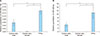
To confirm the microarray results, we identified the expression levels of miR-92a-1-5p, miR-328-3p by real-time polymerase chain reaction (PCR) in FPHL, female and male normal skin groups under study. The analysis showed miR-92a-1-5p and miR-328-3p to be up-regulated that corresponded to microarray results (Fig. 2).
Then we applied DIANA-miRPath v3.0 to recognize signaling pathways regulated in FPHL skin by altered microRNAs. 40 signaling pathways were evaluated as modulated in FPHL versus female control skin, 32 - versus male controls (Supplementary Table 3, 4). Among those, the top pathways revealed their involvement in biological processes regulation such as pluripotency of stem cells, RAS, adherens junction, neurotrophin signaling pathway, estrogen and thyroid hormone signaling pathways.
The analysis of signaling pathways and target genes was carried out for miR-92a-1-5p and miR-328-3p microRNAs (Supplementary Table 5). MiR-92a-1-5p was found to be associated with abnormal fatty acid biosynthesis and metabolism as well as O-glycan biosynthesis and glycan degradation with subsequent gene targets which are presented in the Supplementary Table 3. MiR-328-3p was shown to be implicated in the thyroid hormone synthesis, monosaccharides metabolism, calcium, and intercellular matrix receptor signaling pathways with corresponding target genes.
According to microarray analysis, up-regulations occurred in more than nine hundred of microRNAs in FPHL skin as compared to female or male normal skin. The microarray data were confirmed by real-time PCR analysis. The upper trunk area in healthy males was chosen as it as a sebum-reach and androgen-dependent zones in adults. Our experiment revealed differences in microRNA patterns of healthy female skin from scalp areas, healthy male skin from the back, and FPHL skin. The pathway analysis results showed that these differences are not directly conditioned by androgen metabolism although endocrine alterations are likely to be present as several hormone signaling pathways were found as modulated.
The miR-92a-1-5p up-regulation was observed in FPHL skin. Bioinformatic study on regulation of miR-92a-5p pathway we performed revealed the highest p-values for biosynthesis and metabolism of fatty acids. Women with FPHL are known to have changes in the lipid profile of dyslipidemia type and show significantly higher triglycerides values, total cholesterol values, LDL-C values, and lower HDL-C values versus controls, respectively6. Cholesterol is the precursor of sex hormones which are important in pathogenesis of FPHL in women. Hypoestrogenism which is frequently caused by menopause or ovariectomy, is associated with FPHL in women. The present study demonstrated that estrogen signaling pathway alterations are enriched by up-regulated microRNAs.
The up-regulated miR-328-3p was found to be strongly related to thyroid hormone synthesis. This is in line with the reported action of miR-328-3p on Wnt signaling by targeting its inhibitor SFRP-17 as the effects of Wnt signaling ability to mediate thyroid hormones are well documented. Additionally, thyroid hormones dysfunctions in FPHL patients may influence the FPHL development since these hormones were shown to be suppressed by dihydrotestosterone.
Besides, RAS signaling pathway was in the top of FPHL dysregulated pathways as compared to female and male normal skin. It is well established that RAS abnormal functioning is present in nevus sebaceous and trichilemmoma which contain sebaceous and follicular elements 89. The rapid growth of hair in young skin is associated with HRAS activation in epidermal stem cells10. Therefore, it may be possible that initial alterations on FPHL skin may happen in epithelial stem cells which are patchily present in the skin mostly in hair follicles. Indirect confirmation of this speculation as a pathway associated with stem cells we found as dysregulated in FPHL.
In conclusion, our study determined the microRNAs supposed to be involved in pathologic events regulation and signaling pathways which could play a role in FPHL.
 XML Download