

 PDF
PDF ePub
ePub Citation
 Print
Print


Dear Editor:
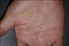
Hypereosinophilic syndrome (HES) is classically defined as (i) persistent eosinophilia of >1,500 eosinophils/mm3 for >6 months; (ii) the absence of any other evident cause of eosinophilia, including allergic diseases and parasitic infections; and (iii) signs or symptoms of organ involvement by eosinophilic infiltration. Skin involvement and cutaneous findings are frequently seen in these patients. Although many other organs other than the skin can also be affected by HES, myopathies associated with HES have rarely been reported1. Here, we report a rare case of focal eosinophilic myositis associated with HES. A 49-year-old woman visited our clinic with a solitary ovoid subcutaneous tender nodule on her right palm that appeared 2 weeks before her visit (Fig. 1). She denied any history of an insect bite or trauma at the site. Routine laboratory tests showed marked elevations in the eosinophil counts (6,730/mm3; reference range, 50~500/mm3), platelet counts (562×103/mm3; reference range, 150~350×103/mm3), and C-reactive protein levels (1.97 mg/dl; reference range, 0~0.6 mg/dl); the other test results were normal. Chest radiography showed mild bilateral pleural effusion. Skin biopsy was then performed, and the patient was referred to the department of allergy to check for the cause of blood eosinophilia. Thorough medical history taking, laboratory examinations, and imaging studies excluded any known causes of hypereosinophilia such as allergic diseases, allergic drug reactions, parasitic infections, human immunodeficiency virus infections, and solid tumors. The skin biopsy showed marked infiltration of eosinophils and lymphocytes in the muscle layer, as well as in the dermis and subcutis (Fig. 2A). The patient later developed a localized erythematous patch on her left calf. The skin biopsy at that site also showed moderate infiltration of eosinophils and lymphocytes in the dermis and subcutis (Fig. 2B). The biopsy specimen was insufficient for the evaluation of the muscle layer. The diagnosis of HES was made. Considering the absence of typical histologic findings of a dermal infiltrate of eosinophils, histiocytes, and eosinophil debris between collagen bundles that form flame figures, a diagnosis of eosinophilic cellulitis was less likely. After treatment with 1 mg/kg methylprednisolone for 2 weeks, the blood eosinophil counts decreased to within the reference range and the skin lesion subsided. The lungs could be another organ involved in HES, taking into account the sudden disappearance of pleural effusion after the treatment.
Recently, to overcome problems with the above-mentioned old definition, Simon et al.2 proposed a new definition for HES: (i) blood eosinophilia (>1,500 eosinophils/mm3) on at least two occasions, or evidence of prominent tissue eosinophilia associated with symptoms and marked blood eosinophilia; (ii) absence of secondary causes of eosinophilia, such as parasitic or viral infections, allergic diseases, drug-induced or chemical-induced eosinophilia, hypoadrenalism, and neoplasm. Our case is consistent with this new proposed definition of HES.

 XML Download