

 PDF
PDF ePub
ePub Citation
 Print
Print


INTRODUCTION
Atopic dermatitis (AD) is a chronic inflammatory skin disease and it has been classified into two different subtypes; classic age-matched subtyping (infantile, childhood, and adult) and presence/absence of allergy (extrinsic AD and intrinsic AD). Lesions can be classified as acute or chronic according to the skin status and differences in cytokine expressions of the lesional skin.
In addition to the presence or absence of sensitization to allergens, AD patients often have impaired skin barrier functions, display significant decreases in the expressions of antimicrobial peptides (AMP) and exhibit innate immune defects, which explain their increased susceptibility to secondary skin infections due to bacteria, fungi or viruses.
Of all the infectious agents found to affect AD patients, the best-characterized is Staphylococcus aureus. The prevalence of S. aureus skin colonization in healthy individuals is 5% to 30%, while 75% to 100% of AD patients have S. aureus on their lesional skin and 30% to 100% of AD patients have this bacteria on their nonlesional skin1-4. A correlation between the severity of eczema and colonization with S. aureus has been demonstrated, which means that bacterial colonization is an important mechanism in the aggravation of skin lesions3,5. Despite the conflicting data, several studies have demonstrated that treatment of S. aureus can decrease the severity of eczema in AD patients6,7. Subjects with severe AD are considered to be more susceptible to S. aureus colonization because they have both innate and adaptive immune defects8.
Th2 polarity with excess production of Th2 cytokines (interleukin [IL]-4, IL-5 and IL-13) may be a key factor in the epithelial production of fibronectin and fibrinogen, which can act as substrates for S. aureus adherence9. There is growing evidence suggesting that this Th2 polarity may adversely affect the innate immune response in the skin of AD patients by inhibiting the productions of AMP and the antimicrobial chemokine MIP3α (CCL20)10,11. Additionally, increased levels of Th2 cytokines inhibit the keratinocyte mobilization of HBD-312. Therefore, biomarkers for Th2 polarity such as serum total immunoglobulin E (IgE) and eosinophil counts can be associated with S. aureus superinfection. Warner et al.13 reported that there was a significantly higher IgE level and greater number of eosinophils in patients with S. aureus colonization than in patients without colonization.
We evaluated laboratory parameters including serum IgE and blood eosinophil counts, and also cultured S. aureus from the skin lesions of AD patients and from the control urticaria patients. We reviewed our data to determine (1) the colonization rates of S. aureus in acute and chronic skin lesions of AD patients and compared them with those of non-AD patients for (2) whether there are differences in the colonization rates according to age, (3) whether Th2 polarity markers (serum total IgE and eosinophil counts) can influence the colonization rates of S. aureus in different age groups and different lesional states.
MATERIALS AND METHODS
Patients
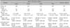
We recruited 687 AD patients (188 infants, 267 children, and 232 adults; 131 acute skin lesions and 556 chronic skin lesions) and 247 control urticaria patients without any skin lesions, including AD or psoriasis, from July 2009 to November 2010. All the patients were examined at the Samsung Medical Center (Seoul, Korea) Dermatology Clinic. Subjects with clinically obvious infections such as impetigo, furuncles or cellulitis were excluded. All the participants did not take any medication during the past 2 weeks. The demographic information is summarized in Table 1.
This study was conducted according to the Declaration of Helsinki, and written informed consent was obtained from all participants. The Samsung Medical Center Ethics Committee approved this study (IRB 2009-12-053-006).
Laboratory markers
Serum total IgE level and blood eosinophil count were measured for all subjects with AD and the control patients. Serum total IgE were measured by Immuno-CAP™ fluorescence enzyme linked assay (Phadia AB, Uppsala, Sweden) and blood eosinophil counts by XE-2100 laser optical flow cytometry (Sysmex Corp., Kobe, Japan). Threshold levels indicative of significances were defined as follows: serum total IgE ≥100 kU L-1 for the infant group, and ≥200 kU L-1 for the child and adult groups, and total eosinophil count ≥300 cells mm-3 for the infant group, and ≥500 cells mm-3 for the child and adult groups.
Isolation and identification of Staphylococcus aureus from atopic dermatitis skin
Subjects with clinically obvious infections, such as impetigo, furuncles or cellulitis, were excluded. We classified the lesions of AD patients into acute and chronic lesions; the acute eczematous lesions were defined by erythema, crusting and oozing; and chronic lesions manifested as papules and lichenification. Bacterial cultures were performed by rubbing rayon-tipped swabs over the lesions (acute skin lesion or antecubital fossa) of the AD patients and on the intertriginous areas of the urticaria patients after cleaning with normal saline. Staphylococcal strains were isolated on blood agar plates and identified as S. aureus using a catalase test and a slide coagulase test.
Statistical analysis
Homogeneity of S. aureus colonization rates across different age groups was analyzed using the χ2 test. Simple or multiple logistic regressions, which was developed to analyze data of binary responses, were used to test for associations of one or multiple factors of diagnostic measurements (serum total IgE level, total eosinophil count and age) and S. aureus colonization rates. We used the significance level of 5% and used SAS (SAS 9.2; SAS Institute, Cary, NC, USA) to conduct the statistical analysis. The 95% confidence intervals of the odds ratios were also used for calculating the results of logistic regression14.
RESULTS
Patterns of Staphylococcus aureus colonization
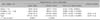
There was a statistically significant difference in S. aureus colonization rates of AD patients and controls with urticaria (p≤0.0001). Colonization rates were 74.0% (97/131) in acute skin lesions of AD patients, 37.9% (211/556) in chronic lesions of AD patients, and 3.2% (8/247) in urticaria patients (Table 2).
When we compared the S. aureus colonization rates in different age groups, we also observed statistically significant differences in both acute and chronic lesions of AD patients (Table 2). The colonization rates in the infant, child, and adult groups were 50.0% (18/36), 80.0% (44/55) and 87.5% (35/40), respectively, for acute skin lesions, and 18.5% (28/151), 41.8% (90/215) and 48.9% (93/190) for chronic skin lesions. This revealed a tendency for the S. aureus colonization rate to increase with age (χ2 test, acute lesions: p=0.0004; chronic lesions: p≤0.0001).
The influences of elevated serum total immunoglobulin E level and the presence of eosinophilia on Staphylococcus aureus colonization rate in atopic dermatitis patients
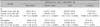
As serum total IgE levels and eosinophil counts have been reported to influence the S. aureus colonization13, we also compared the S. aureus colonization rates according to those parameters. When comparing the colonization results of the elevated serum total IgE group and the normal serum total IgE group, the AD patients in the elevated serum IgE group showed higher S. aureus colonization rates than the normal serum total IgE group with acute and chronic skin lesions. However, the correlation was statistically less significant in acute skin lesions (acute lesion: 81.1% vs. 64.9%, p=0.0387; chronic lesion: 52.8% vs. 23.9%, p≤0.0001). Although the number of S. aureus-positive patients in the urticaria group was small, the elevated total IgE group did not have increased S. aureus colonization rates. Nevertheless, the urticaria patients with elevated total IgE showed decreased S. aureus colonization rates when compared to the normal total IgE group (Table 3A).
When we compared the colonization results according to the eosinophil counts in the chronic lesion group, the patients with eosinophilia showed significantly increased S. aureus colonization rates as compared to those without eosinophilia (52.3% vs. 32.6%, p≤0.0001). However, in the acute lesion and urticaria groups, we did not find any statistically significant differences in S. aureus colonization rates according to the presence of eosinophilia (acute lesion: 71.8% vs. 77.4%, p=0.477; urticaria: 0% vs. 3.4%, p=0.993) (Table 3B).
The combined effects of elevated serum total immunoglobulin E and eosinophilia on Staphylococcus aureus colonization rate in atopic dermatitis patients with acute lesions
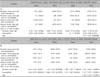
As serum total IgE or eosinophilia alone showed a significant result only in the chronic skin lesion group, we analyzed the combined effects of elevated serum total IgE level and eosinophilia according to S. aureus colonization rate (Table 4). We classified the patients into four groups: the first group had elevated serum total IgE with eosinophilia, the second group had elevated serum total IgE without eosinophilia, the third group had eosinophilia with normal serum IgE, and the fourth group had normal serum IgE and no eosinophilia.
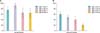
In the acute skin lesion group, the colonization rates of S. aureus of AD patients in the four groups were 75.5% (37/49), 92.0% (23/25), 65.5% (19/29) and 64.3% (18/28), respectively, and there were no statistically significant differences among these groups (Table 4A, Fig. 1A). We also tested these two parameters to the S. aureus colonization rates of AD patients in the different age groups via multiple logistic regressions and found influential effects on S. aureus colonization among the age groups (p=0.00208).
Although we did not detect any statistical significance between these two laboratory parameters and S. aureus colonization in acute skin lesions, we observed a tendency for the S. aureus colonization rate to increase with age.
The combined effects of elevated serum total immunoglobulin E level and eosinophilia on Staphylococcus aureus colonization rate in atopic dermatitis patients with chronic lesions
In chronic skin lesions of AD patients, the colonization rates of S. aureus in the four groups were 60.0% (57/95), 48.9% (81/176), 39.3% (22/56), and 20.0% (46/229), respectively, and there were significant intergroup differences (Table 4B, Fig. 1B). These data revealed that either serum total IgE or eosinophilia can affect the S. aureus colonization as shown in Table 3, and these two parameters showed additive effects on S. aureus colonization in chronic skin lesions of AD patients.
We then compared our results according to age groups. The results for the four groups were 25.0%, 16.7%, 34.1% and 9.5% in infants; 58.0%, 50.8%, 55.6% and 25.8% in children; and 75.8%, 56.0%, 50.0%, and 24.2% in adults. According to these results, only the adult group showed similar results to those of total chronic AD skin lesion.
DISCUSSION
S. aureus is a well-known, Gram-positive bacteria, and the prevalence of S. aureus skin colonization in healthy individuals is 5% to 30%1-4, and those in AD patients is much higher for lesional and nonlesional skin. Acute skin lesions of AD are colonized with greater numbers of S. aureus than the chronic nonlesional skin2, and the density of S. aureus has been shown to be correlated with the degree of cutaneous inflammation and the severity of the AD lesion3. We measured the S. aureus skin colonization rate for Korean AD patients and confirmed that our results were similar to previous data. AD patients with acute skin lesions showed a higher colonization rate (74%) than AD patients with chronic skin lesions (38%). We also found that the S. aureus colonization rates were increased with age in both lesions of AD. A previous study conducted in Sri Lanka showed that the S. aureus colonization rates in acute lesions were higher with ages (47%, <1 year vs. 75%, >15 years)15. However, this study did not observe any differences in the S. aureus colonization rates for chronic lesions among the different age groups.
S. aureus colonization rates in AD patients can be affected not only by lesional state and age, but also by disease severity and duration3,5,15. As nasal S. aureus carriage of caregivers can be a potential source of re-colonization in children with AD16, factors such as school status and family size which can affect the nasal carriage status of S. aureus should also be considered17. In our study, as most of our patients lived in Seoul and suburban areas with high socioeconomic status, we did not take into account the various factors that affect S. aureus colonization except for lesional state and age.
The observed differences in the colonization rates of S. aureus among acute and chronic skin lesions in AD and controls led us to consider the factors affecting the colonization rates of S. aureus and whether the rates may represent an infection or simple colonization of S. aureus in the human skin. Many factors are related to the skin barriers, whether innate and adaptive immune defects have been reported to promote S. aureus colonizations in AD patients. These contributing factors include defective lipid layers, exposed extracellular matrix adhesins, changes in the immune response, bacterial superantigens and increased specific IgE production18. Compared with psoriatic skin lesions and healthy skins, one study found that the expressions of inducible AMP HBD-2 and LL-37 were significantly decreased in AD skin lesions19. The cells of AD patients are unable to efficiently mobilize the HBD-3 to kill S. aureus12. Recently, nonlesional skin of AD patients was shown to have impairments of tight junction proteins, and this may be partially mediated by reduction in the claudin-1 gene20. An IgE reactive protein to S. aureus and S. aureus fibronectin-binding protein (FBP) was identified in the sera of AD patients, and more than one-third of the AD patients showed specific IgE reactivity to FBP21. AD patients have been known to produce IgE-specific antibodies to staphylococcal enterotoxin A or B; however, it was recently reported that S. aureus produces extracellular vesicle (EV) like Gram-negative bacteria22. EV isolated from skin lavage fluids of AD patients contained S. aureus EV-specific proteins, and serum S. aureus EV-specific IgE levels were significantly higher in AD patients than in age-matched controls23.
In this study, we found that the S. aureus colonization rate was higher in the high serum IgE/eosinophilia group as compared to those in the normal IgE/normal eosinophil group in chronic skin lesions but not in acute skin lesions and control group. These two parameters may work additively or synergistically on the colonization of S. aureus, especially in adult patients (Table 4B, Fig. 1B). It might be possible to use these two parameters as predictive markers for S. aureus colonization in chronic skin lesions of AD, however, this result is against previous cytokine reports (Th1 in chronic skin and Th2 in acute skin lesions)24,25.
Our data suggests that, in acute lesions of AD patients, there is a more direct damage to the epidermal skin barrier, mainly through scratching, and S. aureus can easily penetrate or invade the skin and feed off skin exudates. However, in chronic skin lesions of AD, the defects in tight junctions of the lesional skin and S. aureus FBP or S. aureus EV may contribute to the survival of S. aureus in the lesions.
In conclusion, we found that the S. aureus colonization rate was higher in AD patients as compared with non-AD patients, the colonization rate was even higher in acute lesions of AD patients than in chronic lesions, and the S. aureus colonization rate has a tendency to increase with age regardless of lesional status. As the S. aureus colonization rate was higher in the high serum IgE group/eosinophilia group in chronic skin lesions but not in acute skin lesions, we suggest that S. aureus can invade the skin through barrier defects in acute skin lesions of AD. But, the S. aureus colonization in chronic skin lesions of AD patients can be orchestrated through many different factors including tight junction defects in the skin and S. aureus FBP or S. aureus EV formation.
 XML Download