

 PDF
PDF ePub
ePub Citation
 Print
Print


Dear Editor:
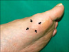
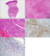
A 61-year-old man presented with several years of history of a solitary cutaneous mass on his left sole. A clinical examination revealed a 26×22 mm sized, skin-colored, firm, asymptomatic subcutaneous nodule (Fig. 1). General laboratory screening, including a complete blood count, renal and liver function tests and lipid levels, were within normal limits. A histopathological examination showed a well-circumscribed mass composed of spindle cells that were arranged in interlacing fascicles or whorls. The spindle cells had elongated nuclei, inconspicuous nucleoli and eosinophilic cytoplasm, resembling smooth muscle cells (Fig. 2). As a result of immunohistochemical staining, these cells were found to be positive for smooth muscle actin, but negative for S-100 protein and desmin. On Masson trichrome staining, many spindle cells with red cytoplasmic stain were found to be dispersed among the blue collagen bundles. Based on both clinical and histological features, a diagnosis of solitary cutaneous myofibroma was made.
Solitary cutaneous myofibroma is a circumscribed benign neoplasm of superficial soft tissue in adolescents and adults; it represents the adult counterpart of infantile myofibromatosis1. Clinically, it typically presents as a painless, slow-growing, firm cutaneous or subcutaneous nodule with an occasional bluish hue. There is a predilection for it to occur on the head and neck, shoulder girdle, lower extremity and hand2,3. Oral and genital solitary cutaneous myofibromas have also been identified. Plantar involvement is exceptionally rare, and there has been only one case of solitary cutaneous myofibroma affecting the sole in the literature3. Histological findings reveal a distinctive appearance, well recognized in children but much less so in adults. It manifests as a biphasic pattern or a zoning arrangement of two cell types4. Among them, the hemangiopericytomatous components, which are typical of infantile myofibromatosis, may sometimes be inconspicuous or even absent in adult lesions, as in our case3. Spindle cells have eosinophilic cytoplasm arranged in short bundles and fascicles resembling leiomyoma. These cells demonstrate features of both myofibroblasts and fibroblasts. Myofibroblastic differentiation of the tumor cells is supported by their immunophenotype. The spindle cells are desmin negative, but smooth muscle actin positive. The Masson trichrome stain, in which thick fibrous bundles with random, irregularly intersecting angles are prominent, can be used to assist in differentiating myofibromas from smooth muscle lesions5. In contrast, smooth muscle lesions show delicate fibrous tissues surrounding the smooth muscle cells and in the septa between the smooth muscle masses. The limited follow-up of solitary cutaneous myofibromas suggests that they tend to follow a benign clinical course with no evidence of recurrence or metastasis1-3.
To our knowledge, our case represents only the second report of a solitary cutaneous myofibroma occurring on the sole. We suggest, even though it is very rare, that solitary cutaneous myofibroma should be considered in the differential diagnosis of solitary lesions involving the sole.
 XML Download