

 PDF
PDF ePub
ePub Citation
 Print
Print


INTRODUCTION
Human papillomavirus (HPV) is identified as an etiological factor in many squamous cell carcinomas (SCCs), particularly of the anogenital area. Recently, SCCs of the periungual area are assumed to have strong relations with HPV infections, for its high HPV detection rates and the existence of concurrent anogenital HPV infections1. In cases of periungual SCCs with coexistent anogenital HPV infections, many mucosal HPV types such as HPV 16, 26, 34, 35, 51, 56 and 58 have been detected in the periungual SCCs1-6. We report a young woman who presented with periungual Bowen's disease after outbreak of intraepithelial neoplasia of the vulva and cervix, and in whom the same mucosal HPVs were identified from these two distant lesions.
CASE REPORT
A 23-year-old woman was referred to us for evaluation of multiple pigmented papules and plaques on the periungual area of both her hands (Fig. 1A). She had a skin-colored verrucous papule on the periungual area of the right thumb for about 8 years. The lesion was previously diagnosed as verruca vulagaris, and then treated by laser vaporization, but not cleared. Four months ago, the lesion became hyperpigmented and enlarged, and spread to the other fingers, especially the periungual area. Three months prior to the changes on her hand, she noticed multiple black plaques on her vulvar area (Fig. 1B). The patient was otherwise well, with no symptoms or signs suggestive of immunodeficiency.
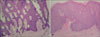
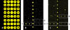
Skin biopsies were performed from the periungual and vulvar lesions. The biopsy specimen from the periungual lesion showed acanthotic epidermis associated with hyperkeratosis. Large hyperchromatic nuclei of dyskeratotic keratinocytes, pyknotic and necrotic cells, and mitoses werescatteredhroughout the epidermis (Fig. 2A). These histological findings were consistent with Bowen's disease. The histology of the vulvar lesion was similar to that of periungual tumors, and the vulvar lesions were diagnosed as bowenoid papulosis of the vulva (Fig. 2B). Moreover, colposcopy disclosed dysplastic change of the cervix, and high-grade squamous intraepithelial lesion was confirmed via a Pap smear. HPV DNA chip and polymerase chain reaction-based microarray system (MyGene CO., Seoul, Korea) were used for the detection and genotyping of HPV from both periungual and vulvar lesion. HPV 11, 18 and 31 were identified from the periungual lesion (Fig. 3B), whereas HPV 11, 18 and 33 were identified from the vulvar lesion (Fig. 3C). The cervical lesion was excised by conization and diagnosed as cervical intraepithelial neoplasia. The periungual lesions were treated with local liquid nitrogen cryotherapy and the vulvar lesions with imiquimod cream. Both lesions regressed but the patient stopped being treated because of personal causes.
DISCUSSION
Bowen's disease is SCC in situ, and is highly associated with sun exposure. Recently, its association with HPV infection has been highlighted1. Although most cases of HPV-associated Bowen's disease occur on the anogenital area, HPV is also identified in approximately 30% of extragenital Bowen's disease2. In HPV-associated extragenital Bowen's disease, the hand is the major site. This can be explained by the fact that the periungual skin is one of the most susceptible sites to this type of infection because the finger and nail unit are vulnerable to trauma1. There is accumulating evidence that periungual Bowen's disease strongly associates with anogenital HPV infections. It tis difficult to calculate the exact concurrent infection rates of periungual Bowen's disease and anogenital cancers, because previous reports have not distinguished periungual Bowen's disease from digital Bowen's disease. However, there is a report in which 7 out of 72 cases of digital Bowen's disease with an antecedent anogenital cancer contain the same HPV subtypes3. According to a recent study, the concurrent anogenital infection rate of periungual SCCs was much higher than that of the proximal or lateral part of finger (0% vs. 100%)1. Furthermore, all HPVs detected from periungual Bowen's disease (HPV 16, 18, 26, 33, 35, 51, 56, 58, 73) belonged to the mucosal types of HPVs, which is thought to be a primary cause of cervical and anogenital cancers4.
The coexistence of the lesions could be best explained by anogenital-digital spread theory5. Viral transmission from the anogenital area to the periungual area is more reliable thanthat in the opposite direction. The reasons are as follows; first, there are higher infection rates of HPVs in anogenital cancer than that of periungual Bowen's disease, second, the time sequence between outbreak of vulvar lesions and ch anges of periungual lesions, and third, the theory of autoinoculation from the anogenital area to the finger by scratching behavior6.
Athough several cases of periungual SCCs with concurrent periungual warts have been reported, it is not certain whether the pre-existent periungual warts changed into periungual SCCs in previous cases. In our patient, there was a solitary, skin-colored papule on her right thumb for 8 years before the changes occurred. Although the pre-existent lesion was not diagnosed by histological tools, it is more reliable to diagnose it as a viral wart because the pre-existent lesion was different from the subsequent lesions in color, size and number of cutaneous lesions. The pre-existed skin colored papule remained unchanged in appearance for several years, but then changed into a hyperpigmented plaque three months after the outbreak of bowenoid papulosis of the vulva. Subsequent hyperpigmented papules developed in other digits after changes of the pre-existed lesion. In the present case, the change from viral wart to SCC possibly occurred by mucosal HPVs from the vulvar lesion, as described above.
It is controversial to perform biopsies on common wartslike lesions in female patients with cervical dysplasia, because only 0.01% of patient who had cervical dysplasia manifested SCC on the finger7. In male patients, the prevalence of digital SCCs among the patients with anogenital disease is not elucidated well. However, if there are evident changes in size, color, or the number of wart-like lesions, as shown in the present case, malignant or precancerous change must be ruled out by skin biopsy. On the other hand, in patients with periungual SCCs or Bowen's disease, careful physical examination of the anogenital area should be performed to investigate dysplasia. Furthermore, the wives of male patients with periungual SCCs also have anogenital dysplasia in some cases1,3. Considering that mucosal HPVs can be transmitted by sexual routes, the efforts to investigate anogenital problems in the sexual partner of patients with periungual SCCs are required.
In summary, patients with periungual Bowen's disease can have a high incidence of concurrent anogenital HPV infection because of viral transmission. Moreover, the presence of HPV within digital SCC is highly related to an increased risk of local recurrence due to the persistence of oncogenic HPV at the margins of the tumor-free plane3. Therefore, careful examination to detect anogenital malignancy should be performed and periodical examination is required for the surveillance of tumor recurrence.

 XML Download