

 PDF
PDF ePub
ePub Citation
 Print
Print


INTRODUCTION
Primary cutaneous lymphoma represents a heterogeneous group of malignancies with clinical findings that are different from those of systemic lymphoma1-3. The recent WHO-European Organization for Research and Treatment of Cancer (EORTC) classification proposed a consensus for a new classification to facilitate more uniformity when diagnosing these cancers1-5. In the new classification, three rare subtypes of primary cutaneous peripheral T cell lymphoma with particular clinical features have been recognized: primary cutaneous CD4 positive small/medium T cell lymphoma (PCSM-TCL), primary cutaneous CD8 positive aggressive epidermotropic cytotoxic T cell lymphoma and primary cutaneous γδ T cell lymphoma2,3. PCSM-TCL usually presents as a solitary plaque or tumor on the trunk of the body and it has a rather favorable prognosis. However, owing to the rarity and heterogeneity of the PCSM-TCL, the precise clinicopathologic characteristics of PCSM-TCL have not been well characterized, and only three such cases have been reported in Korea6-8. We present here an additional rare case of PCSM-TCL that occurred in a 63-year-old woman with multiple, multifocal skin lesions.
CASE REPORT
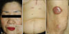
A 63-year-old Korean woman presented with a 5-month history of multiple, multifocal erythematous to violaceous nodules and plaques on the face, abdomen and leg. The skin lesions were intermittently itchy and they had gradually increased in size and number. She had undergone coronary artery bypass graft surgery due to angina in June 2008. Physical examination showed multiple bean-sized erythematous nodules and plaques on the face, leg and abdomen (Fig. 1). There were neither any other systemic symptoms nor lymphadenopathy. The skin biopsy from the thigh demonstrated a dense infiltration in the dermis. The dermal infiltrate showed a proliferation of histiocytes and small/medium-sized atypical lymphocytes with pleomorphism (Fig. 2). On the immunohistochemical studies, most of the infiltrated cells were positively stained with CD3, CD4 and Bcl6, but a few cells were also CD8 and CD20-positive. CD30, CD56 and cytotoxic granules such as granzyme were not seen (Fig. 3). The nuclei of 10% of the tumor cells were stained with Ki-67. Epstein-Barr virus was not detected by in situ hybridization. The staining for terminal deoxynucleotidyl transferase was negative. Monoclonality was demonstrated by means of T-cell receptor γ-chain consensus polymerase chain reaction. No human T-lymphotropic virus type I DNA was detected in the skin biopsy by polymerase chain reaction. The laboratory examinations at the time of the diagnosis, including a complete blood cell with the differential count, liver function tests, renal function tests, an antinuclear antibody test, serum protein electrophoresis and urinalysis were all within the normal limits. The chest X ray, computed tomography of the neck and abdomen and whole body F-18 fluorodeoxyglucose fusion positron emission tomography revealed no abnormalities. The bone marrow aspiration revealed no evidence of malignancy.
She was diagnosed as having primary cutaneous CD4 positive small/medium T cell lymphoma and so electron beam therapy was initiated. The patient received a total dose of 45 Gy for the nasal dorsum in 25 fractions, and 54.2 Gy for the thigh and knee area in 30 fractions twice weekly for 42 days. Approximately 4 weeks after the initiation of treatment, the patient noted flattening of the skin lesion. However, 1 month after completing electron beam therapy, she reported the development of new skin lesions on her back. The skin biopsy specimen from her new skin lesions on the back was consistent with PCSM-TCL. Therefore, she was treated with cyclophosphamide, doxorubicin, vincristine and prednisolone (CHOP) for cutaneous relapse. After the first cycle of chemotherapy, she showed a complete remission of the skin lesions. She is currently being treated with a sixth cycle of CHOP chemotherapy and there is no evidence of local spread or metastasis.
DISCUSSION
PCSM-TCL was included as a provisional entity in the new WHO-EORTC classification for cutaneous lymphomas, and PCSM-TCL makes up 2~3% of all cutaneous lymphomas1-5. This classification of PCSM-TCL is restricted to the cases with predominance of the small/medium-sized pleomorphic CD4 positive T cell phenotype without features of mycosis fungoides1-5.
The majority of patients present with solitary plaques or tumors on the head and neck area or on the trunk. The median age at the onset of the PCSM-TCL was reported to be 60 years9,10. In our case, the age at the onset of the skin lesion was 63 years and she presented with multiple, multifocal lesions. Multifocal lesions similar to our case have been less commonly described11-14.
Histologically, PCSM-TCL is characterized by a dense infiltration of small/medium sized pleomorphic T cells9,11-14. Clonal T cell receptor gene rearrangement has been detected in most cases. Immunohistochemically, CD3 and CD4 are positive, but CD8 and CD30 are negative in the majority of the case, and sometimes this is combined with the loss of the pan T cell markers1,5,11-14. However, many studies have also reported tumors that are CD8 positive or CD4 negative1,5,9,11,12. In addition, many PCSM-TCL cases have shown a rich infiltrate of reactive B cells14. Therefore, the precise clinicopathologic features of PCSM-TCL are not well established and further studies will be needed to define the precise diagnosis and optimal treatment.
The important differential diagnoses are localized mycosis fungoides and T cell pseudolymphoma4,6-8,14. Localized mycosis fungoides is characterized by prominent epidermotropism, which is not present in PCSM-TCL. The other important differential diagnosis is T cell pseudolymphoma, which is characterized by reactive T cell infiltration. It may be difficult to differentiate PCSM-TCL from pseudolymphoma because they demonstrate similar findings such as small lesions with a dense infiltrate of CD8 positive cells and B cells. In this current case, it was helpful to confirm the monoclonality of the T cell receptor gene, which represents true cutaneous lymphomas.
PCSM-TCL has a favorable prognosis with the 5-year survival rate being approximately 60~80%1-4. In previous studies, the overall median survival was significantly better for the patients with localized disease than for the patients with multifocal skin lesions5,15. Furthermore, indolent clinical behavior was associated with stable small lesions (<3 cm), low proliferative activity and an intense CD8 positive lymphocyte infiltration, whereas an aggressive course was associated with rapidly evolving large skin lesions (>5 cm), high levels of proliferation markers and rare CD8 lymphocytes9.
The optimal treatment for this malady has still not been defined4. Localized cases may be treated with topical therapy such as surgical excision, localized psoralen plus UVA (PUVA) treatment bath therapy and local radiotherapy4,6-8,15. In patients with more generalized skin lesions or those who are unresponsive to topical therapy, multi-agent chemotherapy that includes cyclophosphamide may be effective4,5.
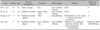
Three Korean cases of PCSM-TCL cases have been reported and only one of the cases presented with multifocal lesions (Table 1). In contrast to the previous reports, our case exhibited an unfavorable course. The patient was treated with multiagent systemic chemotherapy due to the disease progression and cutaneous relapse despite radiotherapy. We think that this may be due to the large, multifocal skin lesions, the scarce CD8 lymphocyte infiltration in the lesion and her relatively old age at the time of the diagnosis.
In summary, we report here on a case of PCSM-TCL in 63-year-old woman with an unfavorable clinical course. PCSM-TCL is a rare heterogeneous disease with various clinicopathologic features that can affect the prognosis. We need to accumulate more cases to facilitate making the diagnosis of this rare entity.


 XML Download