

 PDF
PDF ePub
ePub Citation
 Print
Print


INTRODUCTION
Eruptive vellus hair cysts (EVHC) are characterized by multiple small asymptomatic yellowish or brownish papules with a smooth or centrally umbilicated surface. Their diameters range from 1 to 5 mm. They are usually localized on the chest and proximal extremities, but they may be present at unusual sites, including the face1-10, groin and buttocks10-12. Histologic examination reveals that the cysts are found in the mid-dermis, they are lined by several layers of squamous epithelium and they are filled with cutting vellus hair shafts and laminated keratin materials.
We report here on a case of an EVHC that developed on a rare site, the labium major.
CASE REPORT
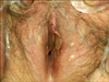
A 64-year-old woman presented with an onset of several yellowish papules on the inner side of her labium major. The clinical examination revealed several well-defined pinhead-to-matchhead-sized yellowish papules that were localized on the inner side of the labium major (Fig. 1). She had felt itching on the both labium majors for 3 years before she visited the hospital. However, the papules on the labium major were asymptomatic and she didn't know when the lesions had first appeared. She had no notable family history of any such occurrences. She had two normal, uncomplicated spontaneous vaginal deliveries, with both pregnancies going full term. A biopsy specimen from the yellowish papule showed a mid-dermal cyst that was lined by a stratified squamous epithelium cells and it was filled with laminated keratin materials and cutting vellus hair shafts (Fig. 2). These findings were consistent with the diagnosis of EVHC.
DISCUSSION
EVHC were first described by Esterly et al.13 in 1977, and they are characterized by multiple small asymptomatic discrete papules that each measure up to 5 mm. This condition does not appear to show a sexual or ethnic predilection, and it is most commonly found in young to middle-aged patients, but it can affect patients at any age14.
The exact pathogenesis of EVHC is unknown, but some theories for it have been proposed. Esterly et al.13 suggested that this condition might arise as a developmental abnormality of the vellus follicles, resulting in occlusion at the infundibular level. This would cause the cyst to retain hairs and keratin. In contrast, Kumakiri et al.2 suggested that the EVHC is a hamartoma of the pilosebaceous unit. Several authors have referred to the close relationship of between the EVHC and steatocystoma multiplex (SM). They suggested that both EVHC and SM may result from a cystic change of the pilosebaceous duct and so they may represent a spectrum of the same clinical entity7,15. Some case reports have described the simultaneous presence of EVHC and SM at one biopsy site3,16,17, or a hybrid cyst showing the combined histopathologic features of EVHC and SM11. However, the cytokeratin studies have shown that the EVHC expresses K17, but not K10, whereas the SM expresses both K17 and K1018, suggesting that EVHC and SM are distinct entities and they are not variants of one disorder.
In most cases, EVHC is found on the anterior chest wall, abdomen and/or extremities, yet other unusual sites can be involved. Kumakiri et al.2 experienced a localized form of EVHC on the forehead and they called it the facial variant, and there has since been several case reports of such facial variants. Less than 15 cases of the so-called facial variant form of EVHC, which is localized exclusively on the face, have been reported1-10. There have also been a few cases on an unusual site such as the buttocks10-12.
The uniqueness of this case is its atypical location. Until now, there have been reports of EVHC on the chest, extremities, face, neck and buttocks, but not on the labium major. To the best of our knowledge, this is first report of eruptive vellus hair cysts on the labium major.
The reason why the lesions were localized only on the labium major in this case is obscure. Some studies have postulated that cystic lesions may be formed by such triggers as minor trauma or scratching, but there is no clear evidence about it. In this case, the scratching for 3 years might have been a triggering factor for the EVHC of the genital area.

 XML Download