

 PDF
PDF ePub
ePub Citation
 Print
Print


INTRODUCTION
Subungual melanoma is frequently misdiagnosed, probably because of its nonspecific clinical features and rarity. Hutchinson1 in 1886 first referred to what he termed1 melanotic whitlow1, a coal-black discoloration at the edge of an inflamed nail. Subungual melanomas represent approximately 2% of all melanomas2 and most often occur in the fifth to seventh decades of life. The common sites reported are the nails of big toes and thumbs. They account for a greater proportion of melanomas in nonwhite persons. One study in a Japanese population found that more than 30% of melanomas involved the digits3.
CASE REPORT
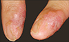
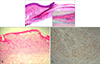
A 36-year-old Korean woman visited our clinic because of a lesion on her right index finger that had been slowly growing over a period of 6 months. On physical examination, 2.5 × 1.5 cm sized erythematous plaque with an incomplete hyperkeratotic surface and partial nail plate loss were observed (Fig. 1). Due to suspicion of Bowen's disease, lichen planus, sarcoidosis, deep mycosis, and others, a punch biopsy was performed. The subsequent histologic examination showed atypical nevoid cell nests which occupied the papillary and reticular dermis. Immunohistochemical staining showed positive reactivity for S-100 protein and HMB-45. There was no sentinel lymph node involvement. Under a diagnosis of amelanotic melanoma of Breslow thickness 2 mm and Clark level IV, she was transferred to our plastic surgery department, where her index finger was amputated below the proximal interpharyngeal joint. A histologic section of the amputated finger showed atypical melanocytic proliferation at the nail matrix dermoepidermal junction. These findings demonstrated that the primary melanoma lesion was located in the nail matrix and that the lesion had spread distally to the nail bed, and proximally to the ventral proximal nail fold, the dorsal proximal nail fold, and to adjacent skin. The tumor cells were ovoid to irregular in shape with slightly hyperchromatic nuclei and moderate cytologic atypia (Fig. 2A). In histologic sections from the nail fold and neighboring finger skin, the tumor was found to be composed of nests with crowded irregular epithelioid cells separated by fibrous septa (Fig. 2B). Immunohistochemical staining for Melan-A showed positive reactivity in melanoma cell cytoplasms (Fig. 2C). Further laboratory examinations, CT and PET CT findings were unremarkable and showed no evidence of metastasis. Her final diagnosis was of amelanotic subungual melanoma, stage Ib.
DISCUSSION
Subungual melanoma is rare, and accounts for only 2 to 3% of all melanoma in light-skinned individuals. However, it represents a significantly higher percentage of melanomas in dark-skinned and Asian people. The fingers are affected more often than the toes. Most tumors occur on the thumb or big toe7. The tumor may present as nail plate loss, a non-healing ulcer, a tumor nodule or subungual pigmentation which often extends into the nail folds78. The incidence of amelanotic melanoma in all melanomas is low, although the subungual region seems to be an area of predilection for amelanotic melanoma in 15 to 25% of cases46. The lack of pigmentation causes the clinical appearance to be nonspecific for melanoma. The differential diagnosis should include a variety of benign and malignant conditions such as subungual hematoma, paronychia, pyogenic granuloma, subungual nevus, subungual fibroma, subungual verruca, keratoacanthoma, Bowen's disease, subungual squamous cell carcinoma, etc59. We considered amelanotic melanoma clinically in our case but also included Bowen's disease, lichen planus, sarcoidosis.
Subungual melanoma is generally associated with poor prognosis, as the majority are deep when diagnosed and early metastases are common. Overall 5-year survival rates range from 20% to 50%. Therefore, early diagnosis is important and subungual melanoma should be considered for all slow-growing and non-healing subungual and periungual lesions, whether it is pigmented or not2310.
 XML Download