

 PDF
PDF ePub
ePub Citation
 Print
Print


Dear Editor:
Incidental focal acantholytic dyskeratosis (IFAD) is a rare incidental histopathological condition characterized by suprabasilar clefts, acantholytic and dyskeratotic epidermal cells, hyperkeratosis, and parakeratosis. Various etiological factors including ultraviolet radiation, hormones, and viral infection have been suggested1. However, the precise etiology of the condition remains unknown. We present the first case of IFAD associated with discoid lupus erythematosus (DLE) and suggest a possible role for the secretory pathway Ca2+ ATPase1 (SPCA1) in the disease.
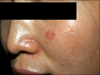
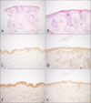
A 38-year-old female presented with a 1-month history of an enlarging, erythematous, pruritic facial plaque (Fig. 1). Laboratory test data including a complete blood count, a liver function test, and measurements of fluorescent anti-nuclear and anti-double-strand DNA antibodies were all normal. Histological examination revealed epidermal atrophy, follicular plugging, loss of the rete ridge pattern, vacuolar degeneration of basal keratinocytes, slight thickening of basement membrane and perivascular and perifollicular infiltration of lymphohistiocytes, compatible with a diagnosis of DLE (Fig. 2A). In addition, a suprabasilar cleft and acantholytic and dyskeratotic cells were also noted (Fig. 2B). The patient was diagnosed with DLE with IFAD and prescribed 0.1% tacrolimus ointment. The lesion had improved moderately at the 1-month follow-up. Patient tissue samples were subjected to further immunohistochemical study, using normal skin from another patient as a control; the tissues were stained with antibodies against the SPCA1 (bs-2434R; Bioss, Woburn, MA, USA) and the sarco/endoplasmic reticulum Ca2+ ATPAase2 (SERCA2) (sc-30110; Santa Cruz Biotech, Dallas, TX, USA). SERCA2 staining was of similar intensity in both tissue samples (Fig. 2C, D). SPCA1 staining of IFAD tissue was less intense than that of normal tissue (Fig. 2E, F).
In 1972, Ackerman2 first described the distinctive histological features of FAD. Changes which had been considered unique to Darier's disease (DD) were also found in a variety of other conditions. FAD presented as either single or multiple lesions. The single lesions were divided into three types: incidental, papular, and nodular12. Various lesions are associated with IFAD; these include epthelial tumors, fibrohistocytic, inflammatory and melanocytic lesions1.
Mutation in ATP2A2 encoding SERCA2 and in ATP2C1 encoding SPCA1 were found in DD and Hailey-Hailey disease (HHD), respectively3. SERCA2 and SPCA1 regulate the Ca2+ levels of the endoplasmic reticulum and Golgi apparatus. Translation, translocation, folding, and processing of adhesional molecule of keratinocyte are closely related with Ca2+ levels; mutations affecting the Ca2+ pumps induce histologically acantholysis and dyskeratosis3. An earlier study revealed that SERCA2 and SPCA1 staining was less intense than normal in both DD and HHD skin, but SERCA2 was more affected in DD patients and SPCA1 in those with HHD45. We found that the SERCA2 staining intensity was similar in both tissues, but SPCA1 expression level was much reduced in IFAD compared to normal tissue. Thus, IFAD seems to be more closely associated with low-level SPCA1 expression.
In summary, to the best of our knowledge, our present IFAD case is the first to be associated with DLE. Although further work is required, our study showed that SPCA1 expression was reduced in IFAD tissue; SPCA1 may thus play a role in disease pathogenesis.
 XML Download