

 PDF
PDF ePub
ePub Citation
 Print
Print


Dear Editor:
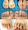
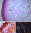
A 54-year-old man presented with a 7-year history of gradually enlarging lesions in the interdigital spaces of both feet (Fig. 1A~D). The lesions were asymptomatic, slightly erythematous, smooth nodules. The patient mentioned repeated mechanical trauma and friction to his feet from working at a construction site. A whole-body skin examination did not reveal any other cutaneous lesions. The biopsy specimen showed large deposits of an eosinophilic, amorphous material located in the entire dermis and subcutaneous fat (Fig. 2A). Amorphous material was also found around the blood vessels and demonstrated apple-green birefringence under polarized light (Fig. 2B, C). A thorough review of systems and a workup were conducted to rule out systemic amyloidosis, but the patient did not report any other abnormalities. Results of complete blood cell count, blood chemistry, urinalysis, serum protein electrophoresis, and urine protein electrophoresis were normal. An abdominal subcutaneous fat pad biopsy failed to demonstrate the presence of amyloid, and an electrocardiogram showed normal left ventricular systolic function and normal myocardial texture. Based on these findings, a diagnosis of primary localized cutaneous nodular amyloidosis (PLCNA) was made. The patient underwent shave excision of the lesions under regional anesthesia. A six-month follow-up showed no signs of local recurrence or systemic progression (Fig. 1E, F).
PLCNA is a rarest form of cutaneous amyloidosis that is defined by the deposition of amyloid fibrils composed of light-chain immunoglobulin in previously normal skin without evidence of deposits in internal organs1. In lichen and macular amyloidosis, the amyloid deposits do not invade blood vessels or extend below the papillary dermis, but the deposition in nodular amyloidosis is found throughout the entire dermis into the subcutis and within the walls of blood vessels1.
Though the pathogenesis of PLCNA is not entirely known, it appears that there is an association between local trauma and deposition of amyloid in nodular amyloidosis, since there have been some case reports of nodular amyloidosis occurring at the sites of prior trauma234. Our patient reported frequent local traumas to the feet during the work, which could have acted as triggers for development of the disease.
PLCNA clinically presents as smooth, yellowish to pinkish nodules ranging from 0.5 cm to several centimeters in diameter1. They are usually located on the extremities, head and trunk. PLCNA occurring on the feet is very rare, with only seven cases reported in the English literature235. PLCNA arising in the interdigital spaces, however, has not yet been reported.
Various methods of treatment such as surgical excision, cryotherapy, and CO2 laser have been employed, but the rate of local recurrence is reported to be high1. Our case of interdigital amyloidosis was successfully treated by simple excision. Appropriate postoperative care is important for preventing complications such as surgical-site infection and wound dehiscence, since interdigital spaces are susceptible to irritant-induced inflammation and bacterial colonization. Ongoing, long-term follow-up is required to detect any sign of local recurrence or systemic changes, since early detection could improve the prognosis of the disease.
 XML Download