

 PDF
PDF ePub
ePub Citation
 Print
Print


Dear Editor:
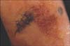
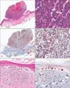
Small cells can be observed in various neoplasms such as small-cell neuroendocrine carcinoma, lymphoma, neuroblastoma, Ewing's sarcoma, and malignant melanoma. Small-cell melanoma, however, is a very rare type of malignant melanoma1. We report a case of malignant melanoma composed mostly of small cells. To our knowledge, this is the first report of such a case in Korea. A 70-year-old female patient was referred to our clinic from a private clinic for an accurate diagnosis of a skin lesion on the right sole. She first recognized the lesion 1 year ago, which seemed to have been increasing in size without any other symptoms. A skin biopsy from the sole was obtained at a private clinic. At the time of her visit, a solitary black patch on sutured skin was noted on her right sole, and irregular brownish to bluish small patches were observed beside the excised area (Fig. 1). The histopathology slide showed a dense aggregation of small cells in the dermis. The cells were small, hyperchromatic, and had scant cytoplasm. On immunohistochemistry, the tumor cells were reactive to HMB-45, and also reactive to S-100 protein and Melan-A. After the lesion was diagnosed as malignant melanoma, the patient was referred to the department of plastic surgery for operation and staging. She was scheduled to undergo surgery 2 months later. During the waiting period, her lesion showed an abruptly enlarged ulcerative mass protruding from the patches. The completely excised mass lesion showed aggregation of dense small cells with vascular invasion into the dermis without lentiginous melanocytic proliferation, whereas the brownish to bluish small patches exhibited lentiginous proliferation of atypical melanocytes (Fig. 2). On computed tomography, metastasis to the liver and bone were noted; her melanoma was diagnosed as stage IV. She was moved to the hematology-oncology department and has since been receiving chemotherapy. The small cell type is a very uncommon subtype, occurring in about 2% of all malignant melanomas2, and manifests as protuberant or ulcerating nodules with aggressive behavior3. Its prognosis, however, is not well documented because of its rarity. Histopathologically, small cell melanoma requires differential diagnosis with other small cell neoplasms, including neuroendocrine carcinoma, lymphoblastic lymphoma, neuroblastoma, and Ewing's sarcoma. In our case, the clinical manifestation of black patch on the sole suggested melanoma. Furthermore, lentiginous epidermal involvement of melanocytes, which was observed as a black patch beside the nodule containing the small cells in our case, is not observed in the other tumor types described above. Although neuroblastoma sometimes stains positive for S-100 protein, positive staining for HMB-45 and Melan-A as well as S-100 protein confirmed the diagnosis of melanoma in our case4. Herein, we report a very rare type of malignant melanoma, small cell melanoma. This case is important because it shows that when aggregation of small cells is observed on histopathology, malignant melanoma should be considered in the differential diagnosis so that patients can receive immediate treatment.
 XML Download