

 PDF
PDF ePub
ePub Citation
 Print
Print


Dear Editor:
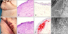
A 58-year-old woman presented with inframammary and inguinal purpuric, non-blanching lesions (Fig. 1A, B).
Eight months before, diagnosis of immunoglobulin A-light-chain plasmocytoma (type lambda) had been made. However, the etiology of skin lesions was still unclear. She did not take any anticoagulants. Prothrombin-time as well as partial-thromboplastin time were normal, and blood count showed only slight thrombocytopenia (110/nl).
An inframammary skin biopsy showed subepidermal amorphous eosinophilic material (Fig. 1C, D) and erythrocyte extravasation. In-situ-hybridization revealed lambda light-chains (Fig. 1F, no kappa light-chains were found: see Fig. 1E); hence, systemic immunoglobulin light-chain amyloidosis was suspected. In electron microscopy, amyloid fibrils were seen (Fig. 1G, H). She received bortezomib/dexamethasone, and it was planned to induce remission for autologous stem cell therapy with lenalidomide/dexamethasone. She died from amyloid-induced heart failure prior to the planned treatment.
Typically, amyloid purpura occurs above the nipple-line, mostly on the head and neck, and particularly on the eyelids1,2. Among the suspected reasons for dermatorrhagia are that factor X is decreased by binding to amyloid fibrils, and that amyloid deposits in blood vessel walls increase vessel fragility.
As purpura may be among the first signs of systemic amyloidoses, it is of utmost importance for dermatologists to keep this sign in mind. A suspected diagnosis of amyloidosis may be the starting point for an interdiscipilinary treatment regimen as different organs may be involved3. Furthermore, it is crucial to treat the underlying cause (e.g., multiple myeloma, plasmocytoma, renal insufficiency with hemodialysis). However, there are also a couple of hereditary systemic amyloidoses with cutaneous involvement, e.g., Meretoja's syndrome (i.e. gelsolin amyloidosis)2. Future treatments with siRNAs or antiamyloid antibodies are in the pipeline4,5, and we will see which ones make their way from bench to bedside.
 XML Download