INTRODUCTION
Kimura's disease (KD) is a rare, chronic allergic inflammatory disorder of unknown etiology1. It is characterized by painless subcutaneous masses, blood and tissue eosinophilia, and markedly elevated serum IgE levels. Although the disease can present at any age, most cases have been reported in the second or third decades of life2,3. Clinically, the subcutaneous soft tissue masses occur predominantly in the head or neck. However, other sites such as the oral cavity, axilla, groin, limbs, and trunk may also be involved1.
We report an unusual case of KD that occurred in a five-year-old boy who presented with a solitary nodule on the left buttock.
CASE REPORT
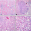
A five-year-old boy presented with an asymptomatic solitary brown pigmented nodule on the left buttock that had been present for several months (Fig. 1). The medical history and family history were unremarkable and there was no report of insect bites or atopic dermatitis. The physical examination revealed left inguinal lymph node enlargement. The laboratory examination revealed markedly elevated serum IgE levels (1,698 ng/ml; NR, ≤100 ng/ml). Other studies including the peripheral eosinophil count, blood urea nitrogen, serum creatinine level, and urinalysis were normal. The histopathology showed a prominent inflammatory cell infiltration, numerous lymphoid follicles with germinal centers, and fibrosis around the lymphoid follicles (Fig. 2A). The lymphoid follicles with germinal centers were surrounded by many eosinophils and proliferating vessels (Fig. 2B). An eosinophilic microabscess was also observed in the reticular dermis (Fig. 2C). The vascular endothelial cells were not epithelioid or histiocytoid, as is usually observed in angiolymphoid hyperplasia with eosinophilia (ALHE) (Fig. 2D). Based on these laboratory and histopathology findings, the patient was diagnosed with KD. The lesion was totally excised and no recurrence has been noted during 15-months of follow-up.
DISCUSSION
KD was first described in China by Kim and Szeto in 1937, and the original description was made by Kimura et al. in Japan in 19481,4. KD is a rare, chronic inflammatory disorder characterized by tumors mainly in the head and neck region, enlarged lymph nodes, increased eosinophil counts, and high serum IgE levels5,6. The disease is most prevalent in Asians, uncommon in Caucasians, and rare in Africans. It has been suggested that one common factor is Asian ancestry3. To date, the etiology of KD remains unclear, though various mechanisms have been proposed. These include: an aberrant immune reaction to C. albicans, parasites, an unknown antigenic stimulus, or localized trauma and clonal rearrangements2,3. Studies have shown that KD is a Th2-mediated disorder with overexpression of interleukin (IL)-4, -5, and mRNAs by the peripheral blood mononuclear cells and lymph nodes7. Type 1 hypersensitivity to some allergens has also been considered as a possible mechanism6.
Although the disease can manifest at any age, most cases have been reported in the second and third decades of life2,3. An earlier review of 194 cases in the Japanese literature revealed that slightly over one-third of the cases occurred in the second decade7. In another case series of 54 Chinese patients, the mean age of the patients was 33.1 years7. The site most frequently affected by KD was the head and neck region (70%)8. Less commonly affected sites often include the groin (15%), extremities (12%), and trunk (3%); rare sites of involvement have included the kidneys, orbits, ears, spermatic cord, and median nerve8. This case presented here has two unusual clinical features: the age of onset (five years old) and the location of the lesion (the buttock). The youngest age reported has been 15 months9, and the buttocks region has never been reported as the location of the lesion.
KD is very similar to ALHE, but is no longer used synonymously. KD is a chronic, allergic inflammatory disease of unknown etiology, whereas ALHE is thought to be a benign vascular proliferative disorder of unknown origin10. Regional lymphadenopathy, peripheral blood eosinophilia, and increased serum IgE levels are commonly seen in KD, but not in ALHE. The nephrotic syndrome due to various types of glomerulonephritis has been more frequently reported in association with KD10. Yuen et al.11 reported that renal involvement is seen in around 60% of cases of KD patients. Histopathologically, KD is characterized by the formation of multiple lymphoid follicles with germinal centers, many of which are infiltrated by eosinophils leading to folliculolysis1,10,12. Eosinophilic infiltration is usually massive, with formation of eosinophilic abscesses10,12. By contrast, in ALHE, the lymphoid infiltration is more diffuse and lymphoid follicles are only occasionally found. Eosinophilic infiltration is more variable10, ranging from sparse to abundant, and eosinophilic abscesses are rare. A vascular component is important in distinguishing KD from ALHE; vascular proliferation of ALHE is more prominent than that of KD. In ALHE, thick-walled blood vessels are lined with hypertrophic, low-cuboidal to dome-shaped epitheloid or histiocytoid endothelial cells containing vacuolated eosinophilic cytoplasm with vesicular nuclei10,12. By contrast, the blood vessels in KD show either flattened or low-cuboidal endothelial cells that do not exhibit the characteristics of the epithelioid or histiocytoid cells10. A summary of the similarities and differences between KD and ALHE is presented in Table 110.
In addition, the clinical manifestations of the case presented here were similar to cutaneous pseudolymphoma caused by insect bites. However, the patient had no history suggesting an insect bite, which may have included a noticeable site of the bite and/or pruritus. Moreover, there was neither epidermal hyperplasia nor wedge-shaped dermal infiltration on the histopathological examination.
The treatment of choice is said to be surgical excision13. Although many alternative treatments, including radiation, high-dose intralesional steroids, and vinblastine have been attempted with good response; however, the tumors usually recur after discontinuing treatment or surgery. Some investigators have reported satisfactory results with cyclosporine7,14 and pentoxifylline13.
The case reported here revealed atypical clinical features of KD. However, the laboratory testing and the histopathological evaluation were consistent with the diagnosis of KD, and not with ALHE, insect bites, or any other malignant neoplasm.


 PDF
PDF ePub
ePub Citation
 Print
Print


 XML Download