

 PDF
PDF ePub
ePub Citation
 Print
Print


INTRODUCTION
Constrictive pericarditis is a disease of the pericardium resulting from chronic inflammation, scarring, or both. In the disease, adhesion of the pericardium to the myocardium limits diastolic expansion of the left and right ventricular walls. The clinical manifestations of constrictive pericarditis include features of left heart failure (pulmonary edema, bilateral pleural effusion, dyspnea on exertion, orthopnea) and right heart failure (ascites, peripheral pitting edema, hepatomegaly). The paradoxical rise in jugular venous pressure or failure to fall on inspiration (Kussmaul sign) in constrictive pericarditis are related to high right atrial pressure and less than full pressure transmission from the pleural cavity to intra-cardiac cavities. The etiologies of pericarditis include post-cardiac surgery, infection, radiation, and malignancy. Because of the increasing amount of cardiac surgery in the modern era, post-operative pericarditis is currently the most common cause of constriction.1)2)3)4)
Irreversibly damaged pericardium with calcification is characteristic of chronic constrictive pericarditis, for which pericardiectomy is the only treatment option.5)6) However, by considering the possibility of early initiating factors and periods of inflammation to the pericardium, detecting and treating early inflammation may be able to halt or even reverse pericarditis. Various cardiac imaging modalities can be helpful to discriminate pericardium with early phase inflammation from chronic inflammation in constrictive pericarditis. In this review, we discuss reversible transient pericarditis and multi-modality diagnostic approaches for transient pericarditis in the context of clinical practice and deciding on an optimal treatment strategy, either surgery or medical treatment.
EVIDENCE OF CHRONICITY
Pericardial calcification is a definitive evidence for chronic constriction with an irreversible scar. Enlargement of both atria in constrictive pericarditis is common due to chronic elevation of diastolic filling pressure in both ventricles. As a result, atrial fibrillation is commonly observed in chronic constrictive pericarditis (30%)7). Advanced liver cirrhosis secondary to cardiac failure can provide further evidence of the chronicity of pericarditis.8)
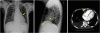
Computed tomography (CT) is the best modality to detect pericardial calcification compared to simple X-ray or echocardiography (Figure 1). A retrospective review of chest CT in constrictive pericarditis showed that CT identifies 68% of cases compared to 49% with chest X-ray.9) The basal aspects of the ventricles and atrioventricular grooves are most frequently calcified (left ventricle n = 23, right ventricle n = 20), followed by the right ventricular outflow tract (n = 20) and right atrium (n = 17). In echocardiography, pericardial calcification frequently distorts the shape of the left ventricle, and myocardial invasion of calcified pericardium can be visualized as high echogenicity with post-echoic shadowing. In chronic constrictive pericarditis, medical therapy with diuretics can improve symptoms in mild cases; however, pericardiectomy is the only way to reverse the constrictive physiology.1)2)10)
TRANSIENT CONSTRICTIVE PERICARDITIS
If constrictive pericarditis is detected during its early stage of inflammation, its disease process can be reversed or improved with/without anti-inflammatory treatment. This condition has been reported as “transient constrictive pericarditis”. Transient constrictive pericarditis was first reported by Permanyer-Miralda et al.11) using M-mode echocardiography. Transient constrictive pericarditis has been subsequently validated as a true clinical entity.12)13)14) One study showed that, among 177 patients with effusive acute pericarditis, 16 had features of constriction, and five had hemodynamic confirmation of constriction by cardiac catheterization, with spontaneous resolution observed in all patients.15) Haley et al.16) reported transient constrictive pericarditis in 36 patients who were treated with anti-inflammatory medication including non-steroidal anti-inflammatory drugs (NSAIDs) and steroids.
The etiologies of transient constrictive pericarditis are diverse. Overall, the most common cause of transient constrictive pericarditis is prior cardiovascular surgery, as for chronic irreversible constrictive pericarditis. However, the causes of acute constrictive pericarditis exhibit regional differences due to the relatively high incidence of tuberculosis in Southeast Asia, including Korea, compared with Western countries.17)18)19) Indeed, tuberculosis is the dominant cause of constrictive pericarditis in Southeast Asia. Other studies (1-5)have suggested that the causes of transient constrictive pericarditis are more variable and include idiopathic causes, postsurgical changes, infection, and connective tissue disease.12)13)14)16)20) A subset of patients in these reports recovered spontaneously, while others recovered after treatment with anti-inflammatory therapy. None of the patients in these studies had radiation-induced pericarditis. Thus, the causes of transient constriction do not appear to differ significantly from those of “classic” constrictive pericarditis.
ROLE OF CARDIOVASCULAR IMAGING IN DIAGNOSIS AND TREATMENT OF TRANSIENT CONSTRICTIVE PERICARDITIS
In the absence chronicity, a diagnosis of transient constrictive pericarditis should be considered. Specifically, transient pericarditis consists of active inflammation of the pericardium and can be treated medically with anti-inflammatory agents. Serial echocardiographic follow-up with Doppler examination can be used to evaluate changes in hemodynamics and physiology in transient constrictive pericarditis (Figure 2).21) Respiratory variation of mitral inflow velocity and tricuspid velocity is augmented in constrictive pericarditis, and diastolic hepatic flow reversal during expiration is a sensitive marker of constrictive physiology. Likewise, interventricular dependence of the left and right ventricles on 2-dimensional echocardiography during the respiratory cycle and changes in pericardial thickness on follow-up may also be useful for assessing improvement of constrictive pericarditis.
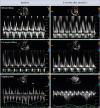
Figure 2
Doppler examination using transthoracic echocardiography is helpful to assess constrictive physiology during treatment. At baseline, respiratory variation of mitral and tricuspid inflow was exaggerated, and diastolic flow reversal during expiration was prominent. After 3 months of steroid therapy, there was no evidence of constrictive physiology.
Serial echocardiographic exams may be sufficient for diagnosing transient constrictive pericarditis. However, a difficulty of clinical decision making is that diagnosis of transient constrictive pericarditis tends to be established during follow-up, after medical treatment is successful or the patient experiences spontaneous recovery. In other words, there are no specific features or markers that can be used to diagnose transient constrictive pericarditis at the time of initial detection of constrictive physiology. Latency of diagnosis may thus result in chronic constrictive pericarditis in patients who would have otherwise experienced disease reversal by anti-inflammatory treatment. Therefore, prediction of reversibility at initial diagnosis of constrictive pericarditis will be helpful to manage the patients.
Inflammatory markers such as C-reactive protein and erythrocyte sedimentation rate tend to be elevated in transient constrictive pericarditis16); however, the power of these biomarkers to predict reversibility is low according to previous reports.19)20)
The reversible nature of transient constrictive pericarditis is thought to be due to relief of active pericardial inflammation in the absence of scarring. Thus, imaging of active inflammation in the pericardium can identify patients with constriction who may respond to a medical therapy. Visualization of inflammation using cardiac magnetic resonance imaging (MRI) based on late-enhancement imaging has been suggested previously.22) In a small pilot study, Feng et al.20) reported that patients with reversible constrictive pericarditis have more intense pericardial inflammation on cardiac MRI and higher inflammatory biomarkers compared to patients without reversibility. Specifically, they reviewed 288 patients who underwent cardiac MRI and identified 89 patients with definite constrictive pericarditis. Among the patients with definite disease, 29 received anti-inflammatory treatment, and resolution of constrictive pericarditis was observed in 14 (48%). In addition, they measured pericardial thickness with late-enhancement and found that the reversible constriction group had increased thickness of late-enhancement (4.4 ± 0.4 mm) compared to the persistent constriction group (2.1 ± 0.4 mm). Thus, detection of pericardial inflammation may be useful, as it may identify patients with transient constrictive pericarditis who are good candidates for anti-inflammatory therapy. Figure 3 shows a typical case of transient constrictive pericarditis with thickened pericardium and constrictive physiology before treatment and recovery to normal physiology and normal pericardium after steroid therapy. Changes in pericardial thickness and disappearance of late-enhancement by the pericardium are also shown in the figure.
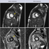
Figure 3
Use of cardiac magnetic resonance imaging to diagnose transient constrictive pericarditis. A 32-year-old man was diagnosed with tuberculous pericarditis. Prior to treatment, the pericardium was thickened (left upper panel) and exhibited signs of late enhancement (left lower panel). After steroid therapy and anti-tuberculous medication, the pericardial thickness normalized and late enhancement of the pericardium resolved.
Positron emission tomography/computed tomography (PET/CT) using 18F-fluorodeoxyglucose (18F-FDG) has been reported as a possible diagnostic tool for predicting the reversibility of pericardial constriction. Indeed, 18F-FDG PET/CT has potential clinical utilization to visualize inflammation with high sensitivity because of high 18F-FDG uptake of active macrophages at the inflammatory site.23) Furthermore, anatomical localization and quantification are possible with 18F-FDG PET/CT.24) Although the degree of 18F-FDG uptake in constrictive pericarditis varies among reports,25) one case report noted significant 18F-FDG uptake by PET in active constrictive pericarditis that disappeared after steroid therapy.26) In addition, Chang et al.19) performed the first prospective study on whether 18F-FDG PET/CT is predictive of response to steroid therapy in constrictive pericarditis. Their study enrolled a total of 16 patients with no evidence of malignancy and no response to short term treatment of NSAIDs or colchicine. All patients were treated with steroid therapy and underwent serial 18F-FDG PET/CT and echocardiography at 3 month intervals with analysis of pericardial SUVmax (maximum standardized uptake value). Nine patients experienced complete recovery from constrictive pericarditis (responder), while 7 patients required either diuretics for symptom control or surgical intervention (non-responder). Pericardial SUVmax at baseline was 7.8 ± 1.4 in responders and 3.1 ± 1.2 in non-responders (p = 0.01). SUVmax was greater than 3.0 in all responders and only in two (29%) non-responders. These results suggest that 18F-FDG PET/CT with a cut-off value of SUVmax = 3.0 may be useful for identifying pericardium with transient constrictive pericarditis. (Figure 4)
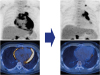
Figure 4
FDG-PET. Maximum-intensity projection (upper row) and fused transaxial PET/CT (lower row) images of a 32-year-old male patient showing significant and diffuse high 18F-FDG uptake in the pericardium (left panel, SUVmax = 16.0). After steroid therapy, the patient was free of symptoms, and 18F-FDG uptake in the pericardium was markedly decreased (right panel, SUVmax = 3.5). CT: computed tomography, FDG: fluorodeoxyglucose, PET: positron emission tomography.
ROLE OF ANTI-INFLAMMATORY TREATMENT
According to a previous study, resolution of symptoms and constrictive physiologic features occurs after a mean of 8.3 weeks.16) The pattern of resolution varies from days to months, and spontaneous recovery is observed in a few cases; however, most patients are treated with an anti-inflammatory agent such as colchicine, NSAIDs, or steroid. Steroid therapy is a frequent treatment option for those not responsive to NSAID or colchicine therapy; however, results of anti-inflammatory treatment need further investigation. Indeed, there have been no randomized controlled studies of anti-inflammatory agents for treatment of constrictive pericarditis. However, in treatment of tuberculosis pericarditis, steroid therapy decreases complications of constrictive pericarditis when applied simultaneous to anti-tuberculous therapy.27)28)
Based on the existing literature, development of chronic constrictive pericarditis appears to begin with acute clinical or subclinical inflammation of the pericardium caused by a variety of mechanical, infectious, iatrogenic, or immunological etiologies. In some patients, the inflammation resolves either spontaneously or with anti-inflammatory medications if they are applied during a curable stage, likely within 2-3 months of the onset of symptoms. If the inflammation does not resolve, the affected tissue will progress toward fibrosis and calcification, eventually leading to clinical heart failure. Inflammatory biomarkers are elevated during an early hyperacute stage when all inflammation can be managed with NSAIDs. As the inflammation becomes subacute, stronger anti-inflammatory agents such as steroids may resolve most pericardial inflammation. In some patients with later stage disease, high doses of steroids may improve residual inflammation, although the fibrotic portion of the pericardium will remain and continue to cause symptomatic constrictive pericarditis. Therefore, in cases of progressive or continuous constrictive pericarditis with evidence of acute inflammation, urgent treatment of the cause of pericarditis combined with optimal anti-inflammatory treatment should be employed to prevent further progression.
CONCLUSIONS
A multi-modality imaging diagnostic approach for constrictive pericarditis is helpful to determine the diagnosis and treatment strategy of constrictive pericarditis. Evaluation of chronicity and reversibility with CT, cardiac MRI, and 18F-FDG PET/CT is useful for establishing a prognosis, as these methods provide reliable evidence as to which patients may improve with anti-inflammatory treatment.
 XML Download