

 PDF
PDF ePub
ePub Citation
 Print
Print


We report a 53-year-old female patient with giant ascending aortic aneurysm accompanied by a severely compressed superior vena cava (SVC) and right pulmonary artery (PA) without dissection.
She presented with NYHA class II dyspnea on exertion and chest heaviness for 2 months and hoarseness of voice for 15 days.
She had no significant family or medical history, no chest trauma and did not smoke. On examination blood pressure was 116/80 mmHg, heart rate 110 bpm and body temperature 36.8°C. All pulses were felt with no radiofemoral or radioradial delay. Jugular venous pressure was not elevated. Oxygen saturation was 98% on room air. Electrocardiogram showed sinus tachycardia, and the chest X-ray revealed an enlarged cardiac silhouette. Hemoglobin was 8.0 g/dL. Other blood tests including erythrocyte sedimentation rate and C-reactive protein, antinuclear antibody, and blood cultures were normal.
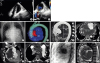
The giant ascending aortic aneurysm was diagnosed on imaging. Transthoracic echocardiography revealed an extracardiac mass compressing the left atrium (Figure 1A-B, Movie 1). Transesophageal echocardiography confirmed the above findings and showed a trileaflet aortic valve. A scanogram of the chest showed lobular mediastinal widening with silhouetting of the ascending aorta, paramedian foci of linear calcification and a laterally displaced prominent conus (Figure 1C). Volume rendered computed tomography (CT) angiogram showed a high thrombus to lumen ratio [Figure 1D, Movie 2 – the smaller patent lumen (600 HU) and a large peripheral thrombus (30 to 60 HU) are color-coded red and blue, respectively]. Multi-detector CT angiography revealed a giant aneurysm (80 × 107× 140 mm) with thrombus and calcification arising from the posterior wall of the ascending aorta sinotubular junction (Figure 1E-F, Movie 3). Severe compression of the SVC (Figure 1G, Movie 4) and right PA (Figure 1H) was seen. Aortography and coronary angiography showed an enormous saccular aneurysm in the posterior wall of the ascending aorta with normal coronaries (Figure 1I, Movie 5, 6, 7, 8). Cardiac MRI showed a variegated hypointense thrombus and severe inferior compression of the atria on sagittal Fast Imaging Employing Steady-state Acquisition (FIESTA) (Figure 1J, Movie 9). An elective surgical procedure was discussed; however, the patient did not consent to the procedure.
Giant ascending aortic aneurysm, defined as an aneurysm with a maximal diameter greater than 10 cm, is rare. Our patient presented with exertional dyspnea, which could be due to anemia and/or the aneurysm compressing the left atrium and adjacent vascular structures. Improvements in multi-modality imaging techniques are helpful in the diagnosis, follow-up, and surgical management planning for ascending aortic aneurysms.

 XML Download