Abstract
Objectives
To describe the incidence and clinical features of adjacent segment disease (ASD) after lumbar fusion and to determine its risk factors.
Summary of Literature Review
The reported incidence of adjacent segment problems is variable, and little has been discussed about surgically treated cases. Risk factors also have not been precisely identified, especially based on structural changes seen on magnetic resonance imaging (MRI).
Materials and Methods
We analyzed the records of 1,124 patients who underwent lumbar or lumbosacral instrumented fusions between August 1995 and March 2006 and had at least one year follow-up. Of these patients, 28 patients who needed secondary operations because of ASD were included in this study. The disease group was compared with an age-, sex-, fusion level-, and follow-up period-matched control group composed of the same number of patients, toward the purpose of analyzing six variables as risk factors.
Results
The incidence of ASD requiring surgical treatment was 2.48%. The mean patient age was 58.4 years, which showed no statistically significant difference from that of the population in which ASD did not develop (57.0 years, p=0.429). Only 1 distal ASD occurred among 21 floating fusions. Facet degeneration was a significant risk factor (p®0.01) on logistic regression analysis.
REFERENCES
1). Shono Y, Kaneda K, Abumi K, McAfee PC, Cunningham BW. Stability of posterior spinal instrumentation and its effects on adjacent motion segments in the lumbosacral spine. Spine. 1998; 23:1550–1558.

2). Bastian L, Lange U, Knop C, Tusch G, Blauth M. Evaluation of the mobility of adjacent segments after posterior thoracolumbar fixation: a biomechanical study. Eur Spine J. 2001; 10:295–300.

3). Park P, Garton HJ, Gala VC, Hoff JT, McGillicuddy JE. Adjacent segment disease after lumbar or lumbosacral fusion: Review of the literature. Spine. 2004; 29:1938–1944.

4). Ha KY, Schendel MJ, Lewis JL, et al. Effect of immobilization and configuration on lumbar adjacent-segment biomechanics. J Spinal Disord. 1993; 6:99–105.

5. Dekutoski MB, Schendel MJ, Ogilvie JW, et al. Comparison of in vivo and in vitro adjacent segment motion after lumbar fusion. Spine. 1994; 19:1745–1751.

7). Cho KJ, Lee JY, Oh IS, et al. Change of segmental motion after lumbar posterolateral fusion. J Korean Orthop Assoc. 1999; 34:281–287.

8). Chung JY, Seo HY, Jung JW. Surgical treatment of adjacent degenerative segment after lumbar fusion. J Korean Spine Surg. 2000; 7:264–270.
9). Kim HT, Jang BD, Hyun KH, Nam JM. Treatment for the sequential degenerative changes at the adjacent segments to lumbar fusion. J Korean Spine Surg. 2000; 7:386–395.
10). Ha KY, Kim YH, Kang KS. Surgery for adjacent segment changes after lumbosacral fusion. J Korean Spine Surg. 2002; 9:332–340.

11). Phillips FM, Carlson GD, Bohlman HH, Hughes SS. Results of surgery for spinal stenosis adjacent to previous lumbar fusion. J Spinal Disrod. 2000; 13:432–437.

12). Chen WJ, Lai PL, Niu CC, Chen LH, Fu TS, Wong CB. Surgical treatment of adjacent instability after lumbar spine fusion. Spine. 2001; 26:519–524.

13). Aiki H, Ohwada O, Kobayashi H, et al. Adjacent segment stenosis after lumbar fusion requiring second operation. J Orthop Sci. 2005; 10:490–495.

14). Brodsky AE, Hendricks RL, Khalil MA, et al. Segmental lumbar spinal fusions. Spine. 1989; 14:447–450.
15). Pfirmann CWA, Metzdorf A, Zanetti M, Hodler J, Boos N. Magnetic resonance classification of lumbar intervertebral disc degeneration. Spine. 2001; 26:1873–1878.

16). Weishaupt DW, Zanetti M, Boos N, Hodler J. MR imaging and CT in osteoarthritis of the lumbar facet joints. Skeletal Radiol. 1999; 28:215–219.

17). Perdriolle R. The torsion meter: a critical review. J Pediatr Orthop. 1991; 11:789.
18). Landis RJ, Koch GG. The measurement of observer agreement for categorical data. Biometrics. 1977; 33:159–174.

19). Ahn DK, Lee S, Jeong KW, Park JS, Cha SK, Park HS. Adjacent segment failure after lumbar spine fusion. J Korean Orthop Assoc. 2005; 40:203–208.
20). Rahm MD, Hall BB. Adjacent-segment degeneration after lumbar fusion with instrumentation: A retrospective study. J Spinal Disord. 1996; 9:392–400.
21). Hambly MF, Wiltse LL, Raghavan N, Schneiderman G, Koenig C. The transition zone above a lumbosacral fusion. Spine. 1998; 23:1785–1792.

22). Wiltse LL, Radecki SE, Biel HM, et al. Comparative study of the incidence and severity of degenerative change in the transition zone after instrumented versus noninstrumented fusions of the lumbar spine. J Spinal Disord. 1999; 12:27–33.
23). Hilibrand AS, Robbins M. Adjacent segment degeneration and adjacent segment disease: the consequences of spinal fusion? Spine J. 2004; 4:190–194.

24). Lehmann TR, Spratt KF, Tozzi JE, et al. Long-term follow-up of lower lumbar fusion patients. Spine. 1987; 12:97–104.

25). Penta M, Sandhu A, Fraser RD. Magnetic resonance imaging assessment of disc degeneration 10 years after anterior lumbar interbody fusion. Spine. 1995; 20:743–747.

26). Okuda S, Iwasaki M, Miyauchi A, Aono H, Morita M, Yamamoto T. Risk factors for adjacent segment degeneration after PLIF. Spine. 2004; 29:1535–1540.

27). Miyakoshi N, Abe E, Shimada Y, Okuyama K, Suzuki T, Sato K. Outcome of one-level posterior lumbar interbody fusion for spondylolisthesis and postoperative intervertebral disc degeneration adjacent to the fusion. Spine. 2000; 25:1837–1842.

28). Aota Y, Kumano K, Hirabayashi S. Postfusion instability at the adjacent segments after rigid pedicle screw fixation for degenerative lumbar spinal disorders. J Spinal Disord. 1995; 8:464–473.

29). Etebar S, Cahill DW. Risk factors for adjacent-segment failure following lumbar fixation with rigid instrumentation for degenerative instability. J Neurosurg. 1999; 90:163–169.

30). Chou WY, Hsu CJ, Chang WN, Wong CY. Adjacent segment degeneration after lumbar spinal posterolateral fusion with instrumentation in elderly patients. Arch Orthop Trauma Surg. 2002; 122:39–43.

31). Ghiselli G, Wang JC, Hsu WK, Dawson EG. L5-S1 segment survivorship and clinical outcome analysis after L4-L5 isolated fusion. Spine. 2003; 28:1275–1280.

32). Gillet P. The fate of the adjacent motion segments after lumbar fusion. J Spinal Disrod Tech. 2003; 162:338–345.

33). Ghiselli G, Wang JC, Bhatia NN, Hsu WK, Dawson EG. Adjacent segment degeneration in the lumbar spine. J Bone Joint Surg Am. 2004; 86:1497–1503.

34). Pellise F, Hernandez A, Vidal X, Minguell J, Martinez C, Villanueva C. Radiologic assessment of all unfused lumbar segments 7.5 years after instrumented posterior spinal fusion. Spine. 2007; 32:574–579.

35). Ha KY, Kim KW, Park SJ, Lee YH. Changes of the adjacent-unfused mobile segment after instrumental lumbar fusion. J Korean Spine Surg. 1998; 5:205–214.
36). Frobin W, Brinckmann P, Kramer M, Hartwig E. Height of lumbar disc measured from radiographs compared with degeneration and height classified from MR images. Eur Radiol. 2001; 11:263–269.
37). Guigui P, Wodecki P, Bizot P, et al. Long-term influence of associated arthrodesis on adjacent segments in the treatment of lumbar stenosis: a series of 127 cases with 9-year follow-up. Rev Chir Orthop Reparatrice Appar Mot. 2000; 86:546–557.
38). Umehara S, Zindrick MR, Patwardhan AG, et al. The biomechanical effect of postoperative hypolordosis in instrumented lumbar fusion on instrumented and adjacent spinal stenosis. Spine. 2000; 25:1617–1624.
Fig. 1.
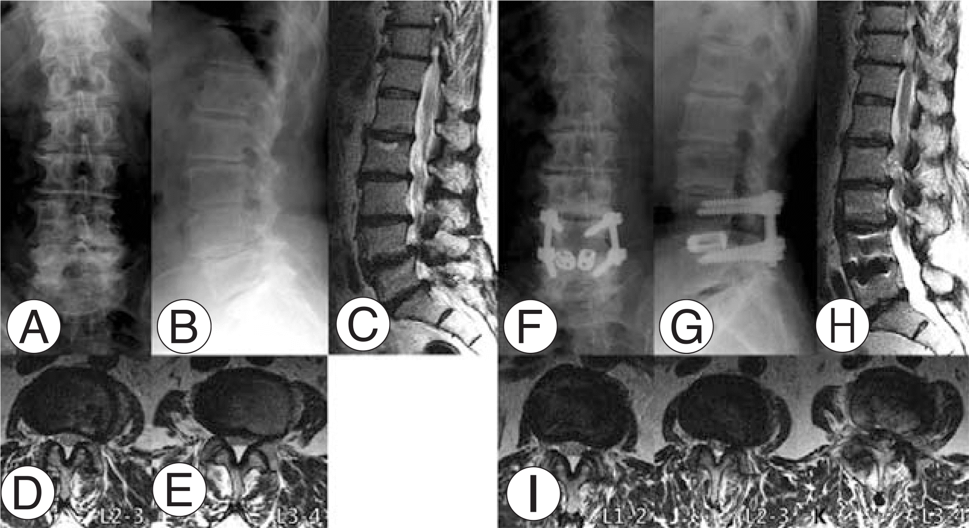
Imaging studies of case 1. She had degenerative spondylolisthesis at L4-5 (A, B and C). Initial radiographs and MRI demonstrate rotational deformity at L3-4, disc wedging at L2-3, and grade 4 disc degeneration at L2-3, L3-4 and L5-S1 (C). Facet degeneration was grade 2 at L2-3 (D) and grade 1 at L3-4 (E). PLIF was performed at L4-5. Adjacent segment disease developed at L2-3 and L3-4 after 33 months (F, G and H). Central spinal stenosis aggravated at L2-3 (J) and L3-4 (K). Facet joints were intact at L1-2 (I). Revision surgery was performed from L2 to L5. Note that L5-S1 segment does not show any deterioration of degeneration.

Table 1.
Classification of disc degeneration15)
Table 2.
Criteria for grading osteoarthritis of the facet joints16)
Table 3.
Patient profiles of disease group.
DSL: Degenerative Spondylolisthesis, SS: Spinal Stenosis, HIVD: Herniated Intervertebral Disc, DS: Degenerative Scoliosis, SL Spondylolysis, RL: Retrolisthesis, ISL: Isthmic Spondylolisthesis, PLF: Posterolateral Fusion, PLIF: Posterior Lumbar Interbod Fusion, ALIF: Anterior Lumbar Interbody Fusion, ASD: Adjacent Segment Disease, AL: Anterolisthesis, LL: Lateral Listhesis VCF: Vertebral Compression Fracture.
Table 4.
Risk factors.


 PDF
PDF ePub
ePub Citation
 Print
Print


 XML Download