

 PDF
PDF ePub
ePub Citation
 Print
Print


Abstract
Objectives
We wanted to determine the effectiveness of performing transpedicular screw fixation in idiopathic scoliosis surgery by evaluating the radiological results.
Literature Review Summary
Deformity correction using pedicle screw fixation in scoliosis surgery is one of the most effective methods of treatment. However, the extent of correction and the complication rate are quite variable.
Materials and Methods
W e evaluated the radiological results of performing posterior correction with using transpedicular screws in sixty patients who were suffering with idiopathic scoliosis. The follow- up duration was 39 months (range: 12 to 91 months). The changes of the coronal and sagittal geometry in the major and compensatory curves were measured according to the Cobb method with using the anteroposterior and lateral radiographs in the standing and lateral bending positions before the surgery and at the final follow- up.
Results
In the coronal plane, the average correction rate of the major curve was 77% and that of the compensatory curve was 72% on the immediate postoperative radiographs. In the sagittal plane, the Cobb angle in those patients who had a hypokyphosis under 15° was improved from 9.0° to 21.2°. The angle between the lowest instrumented vertebra and T10, and also the lumbar lordosis did not change significantly. The correction rate of the rotational deformity of the apical vertebra was 55%, and that of the translation degree was 68%. The correction rate of the translation of the lowest instrumented vertebra was 54% and that of the tilting angle was 77%. No patients had a significant loss of correction in the coronal or sagittal plane at the latest follow- up.
REFERENCES
1). Cotrel Y, Dubousset J. New segmental posterior instrumentation of the spine. Orthop Trans. 1985; 9:118.
2). Cotrel Y, Dubousset J, Guillaumat M. New universal instrumentation in spinal surgery. Clin Orthop. 1988; 227:10–23.

3). Boachie-Adjei O, Bradford D. The Cotrel-Dubousset system-results in spinal reconstruction. Spine. 1991; 16:1155–1160.
4). Denis F. Cotrel-Dubousset instrumentation in the treatment of idiopathic scoliosis. Orthop Clin North Am. 1988; 19:291–311.
5). Labelle H, Dansereau J, Bellefleur C, et al. Comparison between preoperative and postoperative three-dimensional reconstructions of idiopathic scoliosis with the Cotrel-Dubousset procedure. Spine. 1995; 20:2487–2492.
6). Lenke LG, Bridwell KH, Baldus C, Blanke K, Schoenecker PL. Cotrel-Dubousset instrumentation for adolescent idiopathic scoliosis. J Bone Joint Surg Am. 1992; 74:1056–1067.
7). Liljenqvist UR, Halm HF, Link TM. Pedicle screw instrumentation of the thoracic spine in idiopathic scoliosis. Spine. 1997; 22:2239–2245.
8). Luque ER. Segmental spinal instrumentation for correction of scoliosis. Clin Orthop. 1982; 163:192–198.
9). Cundy PJ, Paterson DC, Hillier TM, Sutherland AD, Stephen JP, Foster BK. Cotrel-Dubousset instrumentation and vertebral rotation in adolescent idiopathic scoliosis. J Bone Joint Surg. 1990; 72-B:670–674.
10). Herndon WA, Sullivan JA, Yngve DA, Gross RH, Dreher G. Segmental spinal instrumentation with sublam -inar wires. J Bone Joint Surg. 1987; 69-A:851–859.
11). Labelle H, Dansereau J, Bellefleur C, et al. Perioperative three dimensional correction of idiopathic scoliosis with Cotrel-Dubousset procedure. Spine. 1995; 20:1406–1409.
12). Roy-Camille R, Saillant G, Mazel C. Plating of thoracic, thoracolumbar and lumbar injuries with pedicle screw plates. Clin Orthop. 1986; 17:147–159.
13). Marchesi DG, Aebi M. Pedicle fixation devices in the treatment of adult lumbar scoliosis. Spine. 1992; 17:S304–309.
14). Girardi FP, Boachie-Adjei O, Burke SW, Rawlins BA. Surgical treatment of adolescent idiopathic scoliosis: a comparative study of two segmental instrumentation systems. J Spinal Disord. 2001; 14(1):46–53.
15). Puno RM, Grossfeld SL, Johnson JR, et al. Cotr el -Dubousset instrumentation in idiopathic scoliosis. Spine. 1992; 17:S258–262.
16). Suk SI, Lee CK, Kim WJ, Chung YJ, Park YB. Segmental pedicle screw fixation in the treatment of thoracic idiopathic scoliosis. Spine. 1995; 20:1399–1405.
17). Akbarnia BA, Asher MA, Hess WF, et al. Safety of pedicle screw in pediatric patients with scoliosis and kyphosis. Presented at the annual meeting of the Scoliosis Research Society, Ottawa, Ontario, Canada. 1996.
18). Brown CA, Lenke LG, Bridwell KH, Geideman WM, Hasan SA, Blanke K. Complications of pediatric thoracolumbar and lumbar pedicle screws. Spine. 1998; 23:1566–1571.
19). Suk SI, Kim WJ, Lee SM, Kim JH, Chung ER. Thoracic pedicle screw fixation in spinal deformities. Are they really safe? Spine. 2001; 26:S2049–2057.
20). Kim WJ, Suk SI, Kwon CS, et al. Changes in vertebral rotation following segmental pedicle screw instrumentation and rod derotation in idiopathic thoracic scoliosis: Part I CT evaluation. J Kor Orthop Assoc. 1998; 33:1164–1169.
21). Lee SM, Suk SI, Chung ER. Direct vertebral rotation: a new technique of three-dimensional deformity correction with segmental pedicle screw fixation in adolescent idiopathic scoliosis. Spine. 2004; 29:343–349.
22). Lee SM, Suk SI, Kim JH, et al. Technique of pedicle screw fixation and derotation for the improvement of rotational deformity in scoliosis surgery-Derotation-Screw rotation. J Kor Spine Surg. 2000; 7:527–534.
23). Lenke LG, Bridwell KH, Blanke K, Baldus C, Weston J. Radiographic results of arthrodesis with Cotrel-Dubousset instrumentation for the treatment of adolescent idiopathic scoliosis. A five to ten-year follow-up study. J Bone Joint Surg Am. 1998; 80:807–814.
Fig. 1.
Fourteen-year-old girl with adolescent idiopathic scoliosis (King type I, Lenke type 6 C -). (A, B) Preoperative radiographs show 38° of thoracic and 50° of lumbar curve. Translation of the apical vertebra L1 is 35 mm, and tilt angle of the end vertebra L3 is 24°. (C, D) Postoperative radiographs show that thoracic curve corrected to 8° and lumbar curve corrected to 5°. The apical vertebral translation is improved to 8 mm, and end vertebral tilt angle to 0°. (E, F) At 3 years follow-up radiographs, sagittal and coronal alignment has been well maintained.
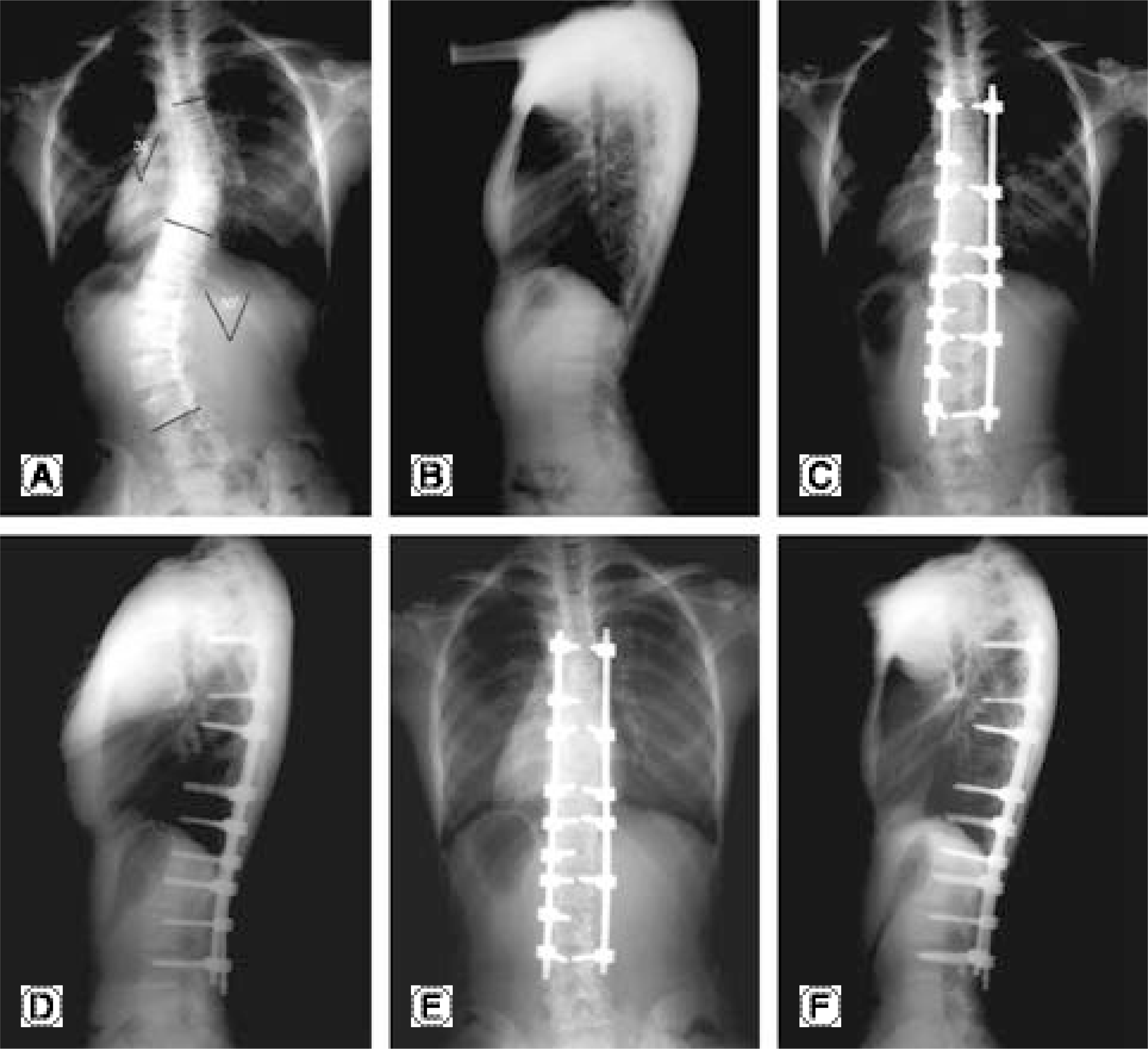
Fig. 2.
Preoperative (A) and postoperative (B) computed tomographs show minimal correction of apical vertebral rotation from 16° to 15°.
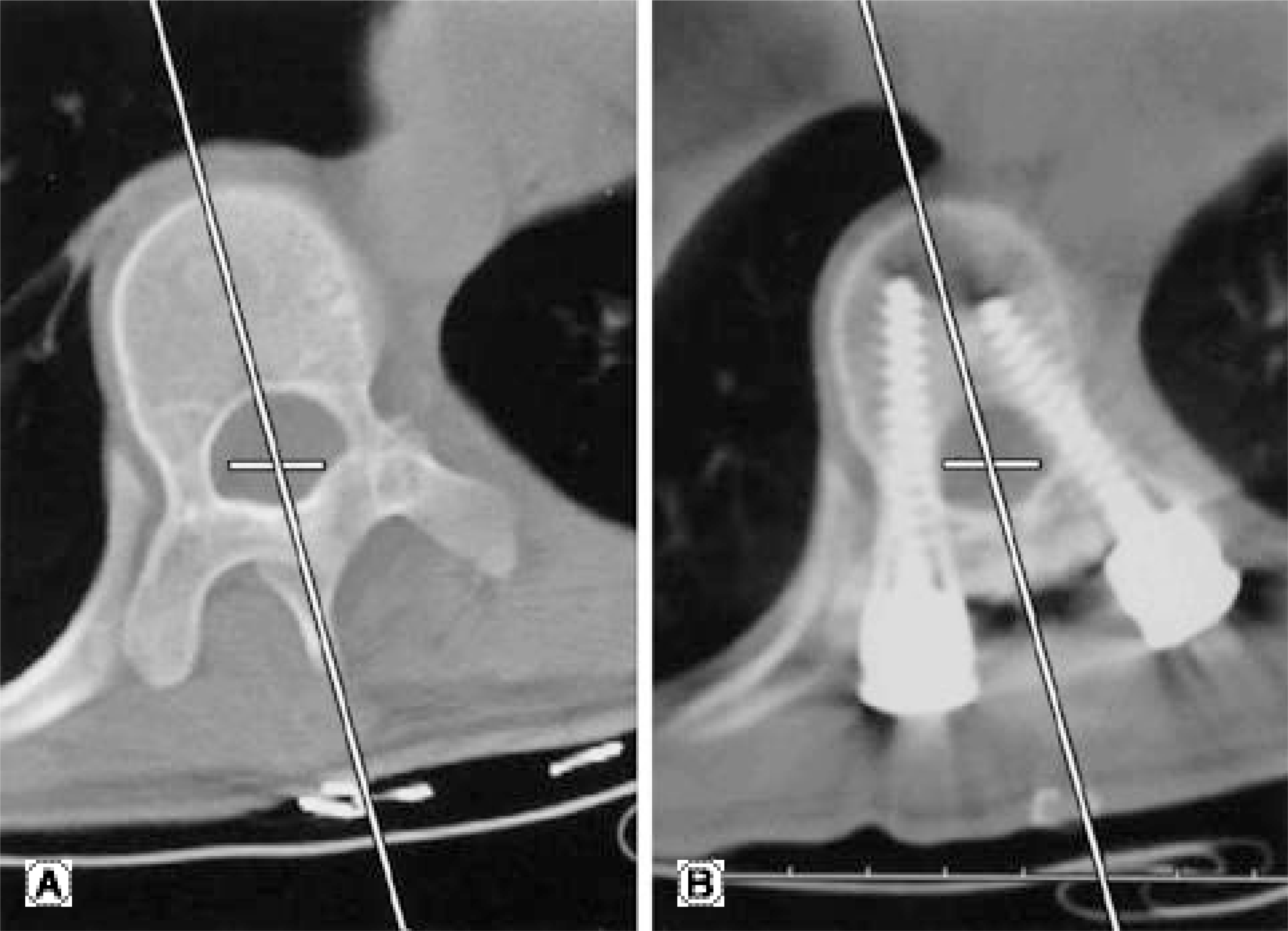
Fig. 3.
A fifteen-year-old girl with adolescent idiopathic scoliosis (King type V, Lenke type 2 B -). (A, B) Preoperative radiographs show 41° of upper thoracic curve and 78° of lower thoracic curve with 75 mm translation of apical vertebra T9. (C, D) Post-operative radiographs show inferior malposition of three pedicle screws (arrows). (E, F) Despite malposition of pedicle screws, radiographs at 2 years follow-up show no loss of correction in sagittal and coronal planes.
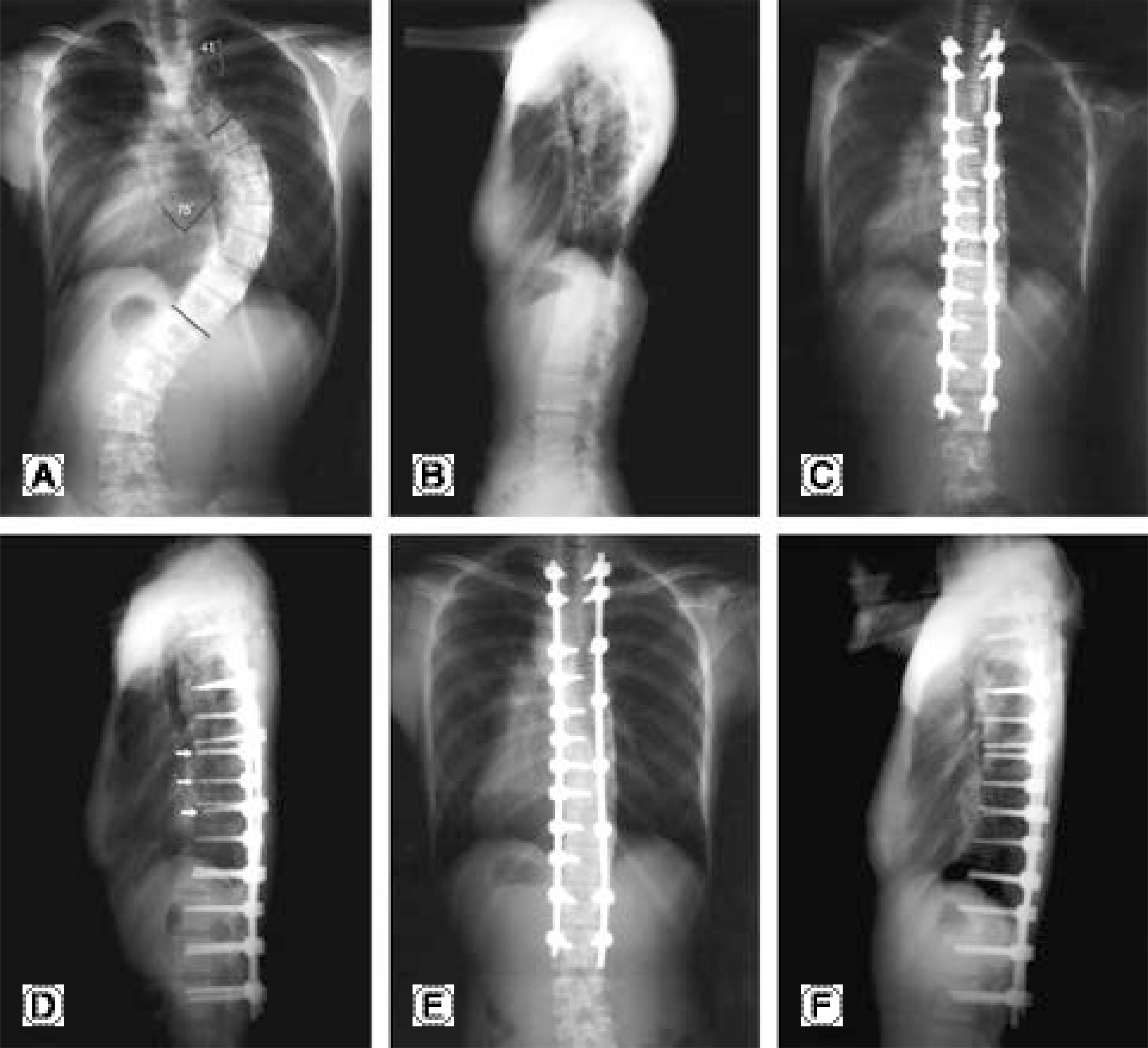
Fig. 4.
Each two consecutive axial images of postoperative CT scan show inferior malposition of pedicle screw (A) and perforation of medial wall of pedicle (B), but neurological complications didn't occur.
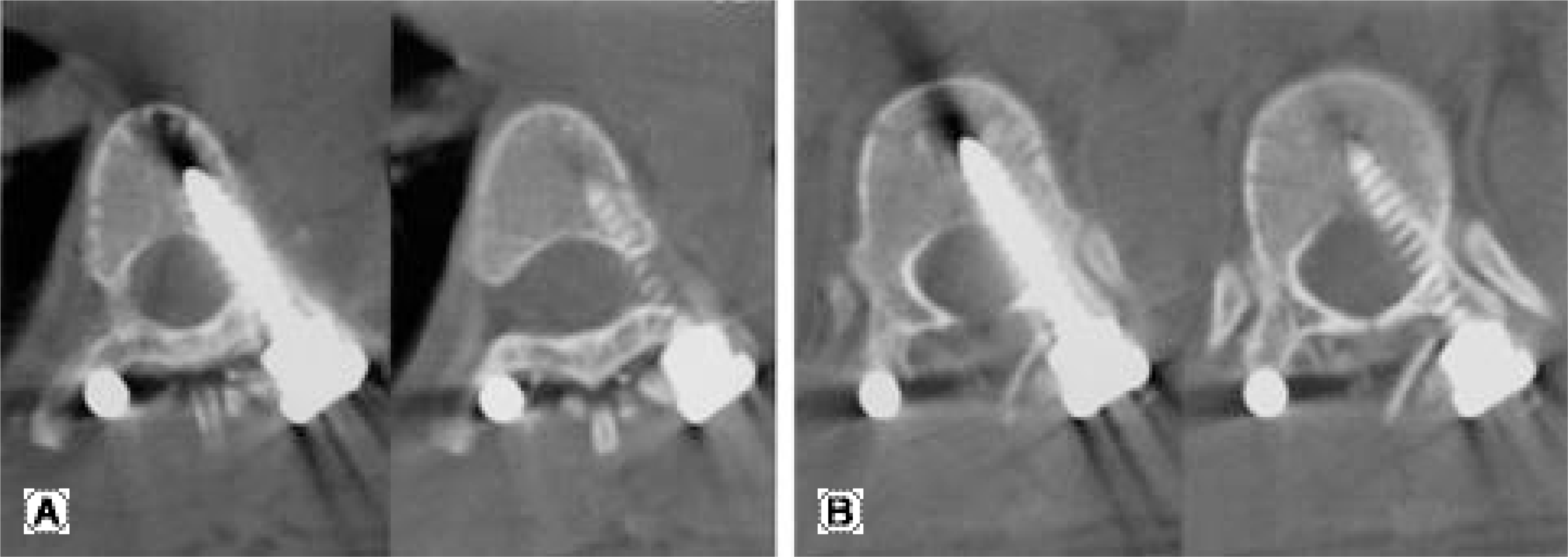
Table 1.
Correction of the primary and compensatory curve
| Preop | Postop | Correction | Follow-up | Correction | Loss of correction | |
|---|---|---|---|---|---|---|
| Primary | 52.0 | 11.9 | 14.3 | |||
| curve | (36∼75) | (3∼20) | 77% | (5∼23) | 72% | 2.4 |
| Compensatory | 30.1 | 8.3 | 10.4 | |||
| curve | (8∼48) | (2∼20) | 72% | (2∼26) | 65% | 2.2 |
Table 2.
Correction of sagittal parameters
| Preop. | Postop. | Follow-up | |
|---|---|---|---|
| T-kyphosis | 19.0 (0-0∼48) | 22.2 (0-5∼37) | 25.2 (-05∼38) |
| T10-LIV∗ | 1.7 (-25∼28) | 0.2 (-27∼18) | 2.3 (-23∼20) |
| L-lordosis | 38.1 (-10∼58) | 36.5 (-18∼60) | 37.6 (-14∼60) |
Table 3.
Correction of parameters in apical vertebra
| Preop | Postop | Correction | Follow-up | Correction | Loss of correction | |
|---|---|---|---|---|---|---|
| Rotation (degree) | 20.6 | 9.5 | 55% | 10.6 | 48% | 1.1 |
| AVT∗ | 38.6 | 12.3 | 68% | 14.6 | 62% | 2.3 |
| (mm) | (8∼82) | (1∼38) | (1∼40) |
 XML Download