Abstract
Objectives
We wanted to identify the changes of ROM and sagittal alignment of the cervical spine after laminoplasty, and we wanted to determine the preoperative factors affecting the ROM and sagittal alignment of the cervical spine after laminoplasty.
Summary of the Literature Review
Cervical laminoplasty is an effective procedure for decompressing multilevel spinal cord compression. It has been reported that the ROM of the cervical spine was decreased after laminoplasty. It is well known that preoperative lordosis of the cervical spine is prerequisite for performing laminoplasty. Maintaining the postoperative lordosis of the cervical spine is also important for decompressing the spinal cord after laminoplasty.
Materials and Methods
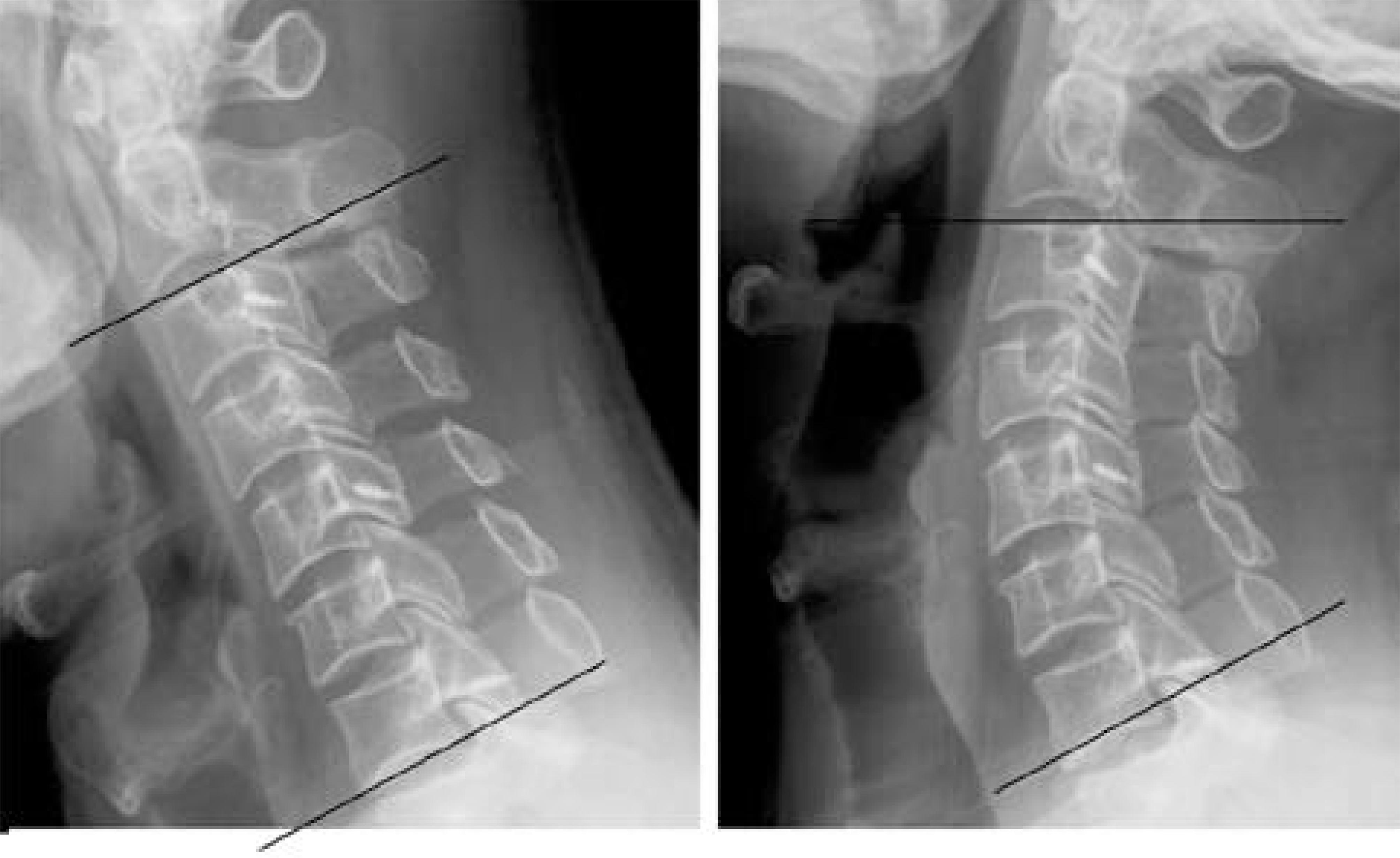
Eighty- five patients who underwent open door laminoplasty from the C3 to C7 levels were prospectively studied. The minimum follow- up was two- years. The preoperative diagnosis was cervical spondylotic myelopathy (CSM) for 52 patients, ossification of the posterior longitudinal ligament (OPLL) for 29 patients and multilevel cervical disc herniation for 4 patients. Plain cervical spine lateral radiography in the neutral, flexion and extension positions was performed preoperatively and at the two- year follow- up. The cervical lordosis or kyphosis was measured by Cobb’s method. The diagnosis, degree of preoperative lordosis in the neutral position, and the degree of preoperative sagittal alignment in flexion and extension were studied as the risk factors for postoperative kyphosis.
Results
The preoperative ROM of the cervical spine was 29.2 degrees and the postoperative ROM was 20.3 degrees. Therefore, 30.5% of the preoperative ROM was decreased after laminoplasty. A decreased ROM of more than 50% was found in 13 patients (15.3%). Their diagnosis was CSM in 11 patients (11/52, 21.1%) and OPLL in 2 patients (2/29, 6.9%). There were no significant differences in preoperative ROM between the two groups with decreased ROM being noted in more than 50% of the patients and decreased ROM being noted in less than 50% of the patients. The preoperative lordotic angle in the neutral position was 16.2 degrees and the postoperative lordotic angle was 11.4 degrees. Kyphosis (mean: 12.2 degrees) developed in 9 patients (9/85, 10.6%) after the surgery. Their preoperative diagnosis was CSM in all patients. The preoperative lordotic angle was significantly less in the kyphotic group than in the lordotic group. The preoperative flexion was 10.2 degrees greater and the preoperative extension was 10.3 degrees less in the kyphotic group than in lordotic group. The preoperative flexion angle was 19.3 degree kyphosis and the extension angle was 8.7 degree lordosis in the kyphotic group.
Conclusions
The ROM of the cervical spine was decreased 30.5% after laminoplasty. Kyphosis developed in 10.6% of the patients. The preoperative factors affecting postoperative kyphosis were the diagnosis of CSM, a preoperative lordosis less than 10 degrees and a greater preoperative flexion angle than the extension angle. Therefore, kyphosis after laminoplasty was expected in a patient with the above three preoperative factors, so other treatment options such as instrumented fusion should be considered.
REFERENCES
2). Sakai Y, Matsuyama Y, Inoue K, Ishiguro N. Postoperative instability after laminoplasty for cervical myelopathy with spondylolisthesis. J Spinal Disord Tech. 2005; 18:1–5.

3). Vatsal DK, Husain M, Jha D, Chawla J. Square cervical laminoplasty incorporating spinous process: surgical technique. Surg Neurol. 2003; 60:131–135.

4). Ishibashi K. Expansive laminoplasty by sagittal splitting of the spinous process for cervical myelopathy: correlation of clinical results with morphological changes in the cervical spine. Kurume Med J. 2000; 47:135–145.

5). Kamioka Y, Yamamoto H, Tani T, Ishida K, Sawamo-to T. P ostoperative instability of cervical OPLL and cervical radiculomyelopathy. Spine. 1989; 14:1177–1183.
6). Wang MY, Shah S, Green BA. Clinical outcomes following cervical laminoplasty for 204 patients with cervical spondylotic myelopathy. Surg Neurol. 2004; 62:487–492.

7). Suda Y, Saitou M, Shioda M, Kohno H, Shibasaki K. Cervical laminoplasty for subaxial lesion in rheumatoid arthritis. J Spinal Disord Tech. 2004; 17:94–101.

8). Suda K, Abumi K, Ito M, Shono Y, Kaneda K, Fujiya M. Local kyphosis reduces surgical outcomes of expansive open-door laminoplasty for cervical spondylotic myelopathy. Spine. 2003; 28:1258–1262.

9). Kawaguchi Y, Kanamori M, Ishihara H, Ohmori K, Nakamura H, Kimura T. Minimum 10-year followup after en bloc cervical laminoplasty. Clin Orthop Relat Res. 2003; 411:129–139.

10). Iwasaki M, Kawaguchi Y, Kimura T, Yonenobu K. Long-term results of expansive laminoplasty for ossification of the posterior longitudinal ligament of the cervical spine: more than 10 years follow up. J Neurosurg. 2002; 96:180–189.

11). Shimamura T, Kato S, Toba T, Yamazaki K, Ehara S. Sagittal splittins laminoplasty for spinal canal enlarge -ment for ossification of the spinal ligaments (OPLL and OLF). Semin Musculoskelet Radiol. 2001; 5:203–206.
Table 1.
Changes of ranges of motion (ROM) after the laminoplasty
| Preoperative | 2-year follow up | P-value∗ | |
|---|---|---|---|
| Flexion (degrees) | -10.2 | -05.6 | 0.000 |
| Extension (degrees) | -19.0 | -14.7 | 0.000 |
| ROM (degrees) | -29.2 | -20.3 | 0.000 |
Table 2.
Comparison of postoperative sagittal alignment depending on preoperative sagittal alignment
| Preop lordosis<10 degrees (n=22) | Postop lordosis>10 degrees (n=63) | P-value∗ | |
|---|---|---|---|
| Cobb’s angle | |||
| at 2-year follow up (degrees) | -0.9 | 15.8 | 0.000 |
| Incidence of kyphosis (%) | 27.2 (6/22) | 4.8 (3/63) |
Table 3.
Comparison of postoperative sagittal alignment depending on preoperative flexion-extension angle
| Flexion > Extension (n=28) | Extension > Flexion (n=57) | P-value∗ | |
|---|---|---|---|
| Cobb’s angle | |||
| at 2-year follow up (degrees) | 1.2 | 16.4 | 0.000 |
| Preoperative lordosis (degrees) | 7.0 | 20.7 | 0.000 |
| Incidence of kyphosis (%) | 25.0 (7/28) | 3.5 (2/57) |
Table 4.
Comparison of preoperative sagittal alignment between kyphotic and lordotic groups
| Kyphotic group (n=9) | Lordotic group(n=76) | P-value∗ | |
|---|---|---|---|
| Cobb’s angle | |||
| at 2-year follow up | -12.2-0 | 14.3 | 0.035 |
| Preop Cobb’s angle in neutral | -09.1 | 17.1 | 0.000 |
| Preop Cobb’s angle in flexion | -19.3-0 | -09.1 | 0.023 |
| Preop Cobb's angle in extension | 8.7 | 20.2 | 0.025 |


 PDF
PDF ePub
ePub Citation
 Print
Print


 XML Download