

 PDF
PDF ePub
ePub Citation
 Print
Print


INTRODUCTION
DNA microarray profiling studies on breast cancer have identified distinct subtypes of this cancer that are associated with different clinical outcomes.(1) Breast cancer was classified into at least four subtypes: luminal A (estrogen receptors (ER) or progesterone receptors (PgR) positive and HER-2 negative), luminal B (ER or PR positive and HER-2 positive), HER2-overexpressing (ER or PgR negative and HER-2 positive), triple negative breast cancer (TNBC) (ER and PR and HER-2 negative).(2,3) The HER2-overexpressing and TNBC had a poor prognosis compared with the luminal subtype. But lack of an established therapeutic target there was no benefit from doxorubicin but the addition of paclitaxel was known to improve both disease-free survival and overall survival.(1-3) But no biomarkers have been identified that can reliably predict a clinical benefit from paclitaxel in women with breast cancer. However few studies have examined whether the CK5/6 positivity can predict a benefit of paclitaxel. Results of preclinical studies and preliminary clinical studies regarding an interaction between CK5/6 and paclitaxel are inconsistent. For these reasons, we investigated whether addition of paclitaxel after adjuvant treatment with doxorubicin plus cyclophosphamide can improve both disease free survival rate and overall survival rate in TNBC. Furthermore we wanted to investigate CK5/6 positivity in TNBC identifies patients with breast cancer who are likely to benefit from the addition of paclitaxel after adjuvant treatment with doxorubicin plus cyclophosphamide.
METHODS
1) Patients
Data were obtained from 616 patients with invasive breast cancer who were diagnosed between 2001 and 2005 at Kosin University Hospital. None of these cancer patients received treatment prior to surgery. We randomly selected 87 women from 104 women with TNBC who had been randomly assigned to receive doxorubicin (60 mg per square meter of body-surface area) plus cyclophosphamide (600 mg per square meter) for four cycles, followed by four cycles of paclitaxel (175 mg per square meter) or more two cycles of doxorubicin plus cyclophosphamide. The patients underwent standard and partial mastectomies with fully resected axillary dissections. Patients were received anthracycline-containing chemotherapy or paclitaxel-adding method after anthracycline-containing chemotherapy if the tumor was node positive. Median follow-up was 5.5 years (range, 0.3~14.8 years), during which there were 15 relapses and 10 deaths. The luminal A/B, HER-2 overexpressing and triple-negative breast cancer phenotypes were identified in 368 (59.8%), 144 (23.4%), and 104 (16.8%) of the 616 cases of invasive breast cancer, respectively. All patients provided written informed consent.
2) Immunohistochemistry
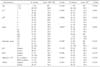
All data were collected from the pathology reports. Histopathological features such as hormone receptor status and HER-2 status on immunohistochemistry (HercepTest, Dako, Denmark) were all analyzed at the Institute of Pathology at the Kosin University. The expression of ER, PgR, HER-2, CK5/6 and other biological markers was determined immunohistochemically in paraffin-embedded tissue specimens. Table 1 summarizes all the antibodies, dilutions, incubation times, and cutoff values used for this analysis. Histological sections (4µm in thickness) were transferred to silane-coated slides and were air-dried overnight at 37℃. The sections were deparaffinized in xylene and rehydrated through a graded series of decreasing ethanol concentrations. Endogenous peroxidase activity was blocked for 10 min in methanol containing 0.3% hydrogen peroxide, and the slides were then rinsed with Trisbuffered saline solution. The expression of ER or PgR was designated as positive when at least 10% of the tumor nuclei showed positive staining. The expression of HER2 was classified according to the HercepTest® assay's scoring system, which includes four categories, namely 0, 1+, 2+ and 3+, based on the intensity and proportion of membrane staining in tumor cells. Positivity was defined as a HER2 score of 3+ for immunostaining or a ≥2-fold increase in HER2 gene amplification, as determined by fluorescence in situ hybridization (FISH). The expression of Ki67 was measured as the percentage of stained nuclei in 1,000 tumor cell nuclei. The measurements were performed in five randomly selected fields, with the Ki67 labeling index determined as the average of these values. The expression of CK5/6, p53 was designated as positive when at least 10% of the tumor cells showed positive staining ER-positive and/or PgR-positive expression combined with HER2-negative expression was defined as HR (+)/ HER2(-). HER2-positive expression, irrespective of the hormone receptor status, was defined as HER2(+). The combination of ER-negative, PgR-negative, and HER2-negative expression was defined as triple-negative breast cancer.
3) Statistical analysis
The primary end point was disease-free survival, defined in the parent study as the interval from study entry until the first local or distant recurrence or death due to any cause. Statistical tests were performed using the SPSS 12.0 statistical software package for Windows (SPSS Inc, Chicago, IL, USA). The survival function was calculated from the time of the onset of disease to the occurrence of death. Survival data were censored on December 31, 2009, which was the date on which the survival data were correlated with the death registry for the last time or 5 years after the onset of the disease. Kaplan-Meier estimates are presented for the survival function, and differences in survival were analyzed using the log rank test. Cox proportional hazards regression analysis was used to estimate hazards ratio and 95% confidence intervals for overall survival and disease-free survival as defined by local recurrence and distant recurrence, whichever occurred first. Associations between specific histopathological and clinical survival estimates and curves were established using the Kaplan-Meier method and differences in observed survival distribution among patient subgroups were tested with two-sided log-rank test. All survival rates were presented with their standard errors. We used Pearson's correlation to determine the association of pairs of explanatory variables and differences in qualitative variables were evaluated by Chi-squared test, where necessary. All P-values were two-sided and a P-value of less than 0.05 was considered to indicate a statistically significant difference.
RESULTS
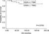
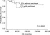
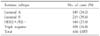
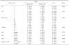
The median age was 46, median tumor size was 3.2 cm. Of the 616 patients, 149 patients (24.2%) were classified as a luminal A type tumor (ER or PR-positive and HER-2-negative), 219 patients (35.6%) were a luminal B type tumor (ER or PR positive and HER-2-positive), 144 patients (23.4%) were a HER-2 type tumor (ER and PR negative and HER-2-positive), and 104 patients (16.8%) were classified as a triple negative type tumor (ER and PR and HER-2-negative) (Table 2). Among patients with triple negative type, 24 patients (27.6%) were classified as CK5/6-positive triple negative type. To verify that each group of selected patients was representative of the CK5/6-positive or negative TNBC population, we compared tumor size, the number of involved lymph nodes, tumor grading, p53, Ki-67, and assigned treatment in the test groups and all treated patients in the trial. There were no detectable differences in clinicopathologic features between CK5/6 positive and negative groups (Table 3). Also among patients with TNBC, 12 patients were classified as paclitaxel chemotherapy groups and 75 patients were classified as no paclitaxel groups. All of the clinical and histopathological data were tested for their prognostic value in a univariate analysis for disease-free survival and overall survival. The 5-year overall survival and disease-free survival probabilities calculated by Kaplan-Meier Estimates (P-value for log rank test) are shown in Table 4. The univariate analysis for prognostic factors associated with disease-free survival in TNBC revealed that the tumor group as T1, T2 or T3 was statistically significant (P=0.034) and that revealed that AJCC staging status as I, IIa, IIb, IIIa, IIIb, IIIc was statistically significant (P=0.0003). The univariate analysis for prognostic factors associated with overall survival revealed that tumor staging was statically significant (P=0.036). Disease-free survival and overall survival among patients who did or did not receive paclitaxel were analyzed according to CK5/6 status in TNBC as established by immunohistochemical analysis (Fig. 1). The apparent interaction between overall survival and the addition of paclitaxel in TNBC group was not significant (P=0.3770) (Fig. 2).
DISCUSSION
TNBCs have been defined as a subgroup of breast cancers that are negative for all ER, PgR, and HER2.(4-6) By using DNA microarray techniques, it has been shown that breast cancers can be classified into biologically distinct groups based on their gene expression profiles.(7-9) These groups comprise luminal A (ER-positive and HER2-negative), luminal B (ER- and HER2-positive), HER-2 (ER-negative and HER2-positive), and triple negative (ER- and HER2-negative) subtypes.(10,11) The TNBC is a heterogeneous group and is further categorized into the basal-like and the normal breast subtypes, which are positive and negative, respectively, for myoepithelial/basal markers such as basal cytokeratins (CKs) (i.e., CK5/6, CK14, and CK17), a-smooth muscle actin, and epidermal growth factor receptor (EGFR).(12,13) Although TNBCs account for only 10~17% of all breast carcinomas, this subgroup is regarded as important clinically because of the aggressive clinical behavior, poorer patient prognosis, and lack of an established therapeutic target.(13-15) In fact, the consensus criteria for the basal-like subtype have not yet been established. Nielsen et al.(8) suggested four representative surrogate markers for the basal-like subtype: ER, HER2, EGFR, and CK5/6. They reported that the sensitivity and specificity of the combination of ER negativity, HER2 negativity, and EGFR and/or CK5/6 positivity were 76 and 100%, respectively.(16) Other additional criteria used for the basal-like subtype comprise ER negativity and HER2 negativity, and vimentin, EGFR, CK8/18, and/or CK5/6 positivity, and triple negativity, and CK5/6 and/or EGFR positivity. Other markers that have been included in the panel of myoepithelial/basal biomarkers are laminin, c-KIT, p63, nestin, osteonectin, caveolin 1, and nerve growth factor receptor.(17-19) In the clinical studies to examine the efficacy of taxane based adjuvant chemotherapy to node positive breast cancers, taxanes (paclitaxel and docetaxel) were shown to significantly improve the prognosis of patients with TNBC. Liedtke et al. compared response to neoadjuvant therapy and long-term survival in 255 patients with stage I~III TNBC and 863 patients with stage I~III non-TNBC.(4,19) They concluded that TNBC compared with non-TNBC had significantly higher rates of pathologic complete response (pCR) (22% vs. 11%, P=0.034).(20,21) Three year disease-free survival rates (63% vs. 76%) and overall survival rates (74% vs. 89%) were also significantly lower in patients with TNBC than in those with non-TNBC. Patients with TNBC with residual disease had significantly decreased overall survival compared with those with non-TNBC with residual disease. In 92 patients who had received neoadjuvant chemotherapy against invasive TNBC in the National Cancer Center Hospital (NCCH), pCR and near pCR (Grade 2b) were acquired in 29 (32%) and 6 patients (6.5%), respectively, and these rates in TNBCs were higher than those in non-TNBCs.(22) The ratio of basal-like subtype in TNBC was estimated to be up to 56~84%. Therefore, characteristic histopathological features of TNBCs are similar with those of the basal-like subtype. Characteristic histopathological types that constitute TNBCs and basal-like subtype are high-grade invasive ductal carcinoma of no special type, typical medullary carcinoma, metaplastic carcinomas, and adenoid cystic carcinoma.(23) Although taxanes were shown to be effective for TNBCs in clinical trials, there are a number of cases resistant to taxanes. It has been suggested that BRCA1 downregulation gives rise to the defects in DNA repair, especially in homologous recombination, and BRCA1 dysfunction confers enhanced sensitivity to certain cross-linking agents, such as platinum salts-based chemotherapeutic agents. Furthermore, it now has been shown that the basal-like subtype comprises heterogeneous groups. At present, 30% of TNBCs are sensitive to standard anthracycline-taxane-based regimens, but it is impossible to predict efficiently the chemosensitive TNBCs histopathologically or by specific biomarkers. For the identification of new targets of molecular therapies and of chemosensitive TNBCs, further research is necessary.
CONCLUSION
We observed an interaction between the CK5/6 status of the TNBC and the benefit of adjuvant paclitaxel in TNBC who received four cycles of doxorubicin plus cyclophosphamide. Our results indicate that CK5/6 positivity can not predict improvement in disease-free survival and overall survival by the addition of paclitaxel to doxorubicin plus cyclophosphamide. But because our study is small size study, more abundant patients' data will be needed to evaluate of the CK5/6's predictive role.

 XML Download