

 PDF
PDF ePub
ePub Citation
 Print
Print


INTRODUCTION
Maternal prenatal stress affects the offspring's risk of noncommunicable diseases (NCDs),1 including allergic diseases.23 Oxidative stress is thought to play a key mechanism by which the prenatal maternal stress affects childhood adverse health outcomes.1 Oxidative stress increases when subjects are under psychological stress, elevating the risk of contracting NCDs.4 The increased oxidative stress is also involved in the pathophysiology of childhood atopic dermatitis (AD).5
Subjects' exposure to oxidative stress cannot be easily quantified. Although the concentration of glutathione (GSH) or the ratio of GSH/oxidized GSH (GSSG) can represent the exposure to oxidative stress,6 the target specimens are hard to acquire. Recent studies proclaimed that telomeres shorten with progressive cell divisions,7 and the shortening of leukocyte telomere length (LTL) reflects the exposure to cumulative oxidative stress.89
The authors aimed to verify the LTL as a candidate biomarker for AD development as well as exposure to stress. In other words, we hypothesized that the LTL would be shortened in offspring following the exposure to prenatal maternal stress, and the LTL shortening could also be associated with AD development during early life. Therefore, we attemted to analyze blood samples archived in a birth cohort regarding their association with the exposure to prenatal maternal stress and with the AD development.
MATERIALS AND METHODS
Subjects
We selected a birth cohort (the COhort for Childhood Origin of Asthma and allergic diseases [COCOA]) that had displayed an epidemiologic association between prenatal maternal stress and the offspring's AD development.1011 It archives the cord blood- and 1-year-old peripheral blood samples. In the COCOA study, prenatal maternal stress was evaluated using 2 self-reported questionnaires at 36 weeks' gestation, namely the Center for Epidemiological Studies-Depression (CESD) 10 and the State-Trait Anxiety Inventory (STAI)-Trait subscale, which measures depression and anxiety, respectively.1213
From the pre-defined 4 distinctive sampling pools, among subjects whose cord blood and 1-year-old peripheral blood were both available, we selected 4 groups of 68 subject samples. Four groups are determined by their exposure to prenatal maternal stress (high or low stress) and their later development of AD up to their scheduled visit at the age of 1 (with later AD or without AD development). As a result, 4 groups are labeled as the high stress with later AD (HSWD), high stress without AD (HSOD), low stress with later AD (LSWD) and low stress without AD (LSOD) groups.
To show the different characteristics between the groups, the low-stress group pools included subjects whose both CESD and STAI scores were in the lowest tertile. Whereas the high-stress group pools included subjects whose both CESD and STAI scores were in the highest tertile or those whose either CESD or STAI score was in the highest quartile. The other subjects were not considered the sampling candidate for this analysis. Subjects' AD development was ascertained by pediatric allergy specialists who made a clinical diagnosis after a detailed history and physical examination based on the criteria of Hanifin and Rajka up to their 1-year scheduled visit.14
Because of the insufficient number of subjects whose both cord-blood and 1-year peripheral-blood samples were available, we measured LTLs from all available 23 subjects in the HSWD group, randomly-selected 20 subjects in the LSOD group, and randomly selected 15 subjects in both the HSOD and LSWD groups, respectively. After we exclude subjects whose cord-blood LTL or 1-year peripheral-blood ones was not successfully measured, we finalized 68 subjects (HSWD, 22 subjects; HSOD, 14 subjects; LSWD, 13 subjects; and LSOD, 19 subjects) as the population for analysis.
From the archived data in the COCOA cohort, 68 subjects' parameters regarding clinical characteristics were obtained. They include the child's sex, mode of delivery, gestational age and birth season. If subjects were born from March to May, they were regarded as being born in spring. From June to August, September to November and December to February were considered the summer, autumn and winter, respectively. Finally, maternal allergy history as well as the maternal and paternal age was also obtained.
The study protocol was approved by the Institutional Review Board of Seoul National University Hospital/Seoul National University College of Medicine (IRB No. 1701-006-819). Since subjects who provided research samples had agreed to secondary use in the original birth cohort, the requirement for written informed consent was waived.
LTL measurement
LTLs were determined from the average terminal restriction fragment (TRF) length, which was measured using a chemiluminescence technique according to the manufacturer's manual of a commercially available Telo TAGGG telomere length assay kit (Roche-Applied Science, Mannheim, Germany).1516 DNA was extracted from the buffy coat of cord blood and 1-year-old peripheral blood using a DNA extraction kit (Qiagen, Crawley, UK). DNA samples (75 ng/mL) were digested with 10 U Rsa I and 10 U Hinf I for 2 hours at 37°C. DNA fragments were separated by electrophoresis at 150 V for 2 hours on 0.8% agarose gels. The DNA samples were then purified with 0.25 M HCl, denatured with NaOH-NaCl (0.5-1.5 M) and neutralized with Tris-NaCl (0.5-3 M, pH 7.5). According to the manufacturer's protocol, DNA samples were transferred onto a positively charged nylon membrane and fixed with Ultraviolet light for Southern blot analysis. Membranes were hybridized with a telomere repeat-specific digoxigenin (DIG)-labeled probe overnight at 42°C and washed thrice with 2× saline sodium citrate (SSC)/0.1% sodium dodecyl sulfate (SDS), followed by 0.2 × SSC/0.1% SDS.
Next, membranes were incubated with a DIG-specific antibody covalently coupled to alkaline phosphatase. Finally, the immobilized telomere probe was visualized using alkaline phosphatase-metabolizing CDP-Star® (Tropix Inc., Bedford, MA, USA). The telomere smear was obtained by printing the membrane on an autoradiograph film (Lumi-Film chemiluminescent detection film; Roche-Applied Science) and scanning using a densitometer. The mean TRF (an estimate of telomere length) of each sample was calculated using the following formula:
where ODi is the optical density at a given position in the lane, and Li is the length in kilobase pairs at that position.
Statistical analysis
Data were analyzed using statistical software (SPSS ver 23.0; IBM Corp., Armonk, NY, USA) and R statistical software (version 3.3.2; R Foundation, Vienna, Austria). Changes in LTL during the first year of life were assessed by the paired t tests, and each group's yearly change was assessed using the Wilcoxon signed rank test. Differences in cord blood and 1-year-old peripheral blood LTLs were assessed using the Mann-Whitney U test, and the absolute amount of attrition in LTLs across all 4 groups was evaluated using the Kruskal-Wallis test followed by post hoc Dunn's test with further P value adjustment by the Benjamini-Hochberg false discovery rate method.17 The proportion of later AD development according to the stress exposure and telomere length was assessed by the Fischer's exact test. The cutoff for shorter LTL was defined by the median of total subjects enrolled in this study. Differences were considered statistically significant when the P value was less than 0.05.
RESULTS
The distribution of subjects' clinical characteristics among the 4 sample groups is listed in Table 1. These all 4 groups were not different in their maternal and paternal age, gestational age, sex ratio, birth weight or delivery type. On the other hand, the ratio of subjects with a maternal allergy history was different between the 4 groups with the lowest percentage in the LSOD group (P = 0.030). However, most of the maternal histories were about the allergic rhinitis (n = 16) and history of AD was only 3 cases (2 and 1 cases in the LSWD and HSWD group, respectively). The distribution of birth seasons was also different between groups without showing any specific pattern (P = 0.041).
Table 1
Distribution of the clinical characteristics among the 4 groups
All variables are presented as the number (%) or the median (IQR).
HSWD, high stress with later atopic dermatitis; HSOD, high stress without atopic dermatitis development; LSWD, low stress with later atopic dermatitis; LSOD, low stress without atopic dermatitis development; AD, atopic dermatitis; CESD-10, Center for Epidemiological Studies-Depression 10; IQR, interquartile range; STAI-T, State-Trait Anxiety Inventory-Trait subscale.
*One subject had maternal history of both asthma and allergic rhinitis concomitantly.
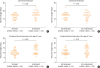
The mean LTL from all cord blood samples was 14.4 ± 3.9 kbp. Fig. 1 showed the difference of cord-blood or 1 year-peripheral blood LTLs according to prenatal stress or later AD development. The cord-blood LTLs were shorter in high-stress groups than in low-stress ones (Mann-Whitney U test; median, 15.0 [interquartile range; IQR, 10.3, 15.0] vs. 15.0 [12.8, 18.0] kbp, P = 0.026; Fig. 1A), but they were not different between groups that later developed AD and those did not (median, 15.0 [IQR, 11.0, 16.0] vs. 15.0 [11.3, 18.0] kbp, P = 0.915; Fig. 1B). On the other hand, the median LTL from all 1-year-old peripheral blood samples was 11.5 ± 3.6 kbp, which was significantly shorter than that of cord blood samples (paired t test, P < 0.001). The 1-year-old peripheral-blood LTLs were still shorter in high-stress groups than in the low-stress ones (Mann-Whitney U test; median, 10.0 [IQR, 8.6, 12.0] vs. 13.0 [9.0, 17.0] kbp, P = 0.008; Fig. 1C), but they were not different between groups that later developed AD and those did not (median, 10.0 [IQR, 8.6, 13.0] vs. 12.0 [8.6, 16.5] kbp, P = 0.174; Fig. 1D).
Fig. 1
Comparison of the telomere length of cord blood leukocytes according to (A) exposure to prenatal stress and (B) AD development. The telomere lengths from 1-year-old peripheral blood were also compared according to (C) exposure to prenatal stress and (D) AD at the age of 1 year.
AD, atopic dermatitis; HSWD, high stress with later atopic dermatitis; HSOD, high stress without atopic dermatitis development; LSWD, low stress with later atopic dermatitis; LSOD, low stress without atopic dermatitis development.
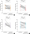
The changes in each subject' LTL during their first year of life between the stress/AD groups are depicted in Fig. 2. Either prenatally stressed or later AD-developed groups presented a significant decrease in LTL during the year (Wilcoxon signed rank test; cord-blood vs. 1-year-old peripheral blood; HSWD group: median, 13.5 [IQR, 10.0, 15.25] vs. 10.0 [8.6, 12.25] kbp, P = 0.005, Fig. 2A; HSOD group: median, 15.0 [IQR, 12.75, 15.75] vs. 10.0 [7.3, 12.0] kbp, P = 0.004, Fig. 2B; and LSWD group: median, 16.0 [IQR, 15.0, 18.0] vs. 10.0 [8.8, 13.0] kbp, P = 0.003, Fig. 2C). On the other hand, those in neither prenatally stressed nor later AD-developed group did not show any significant change (median, 15.0 [IQR, 10.0, 19.0] vs. 15.0 [10.0, 17.0] kbp, P = 0.434; Fig. 2D).
Fig. 2
Changes in telomere length between cord blood and 1-year-old peripheral blood leukocytes from subjects in the (A) HSWD, (B) HSOD, (C) LSWD and (D) LSOD groups. HSWD, high stress with later atopic dermatitis; HSOD, high stress without atopic dermatitis development; LSWD, low stress with later atopic dermatitis; LSOD, low stress without atopic dermatitis development.
LTLs of 1-year-peripheral blood and their changes during their first year of life according to subject groups were displayed in Fig. 3. The 1-year median LTL in the LSOD group was 15.0 [IQR, 10.0, 17.0] kbp, which is the longest among the 4 subject groups (Kruskal-Wallis test, P = 0.016; Fig. 3A). In post hoc analysis, 1-year-peripheral blood LTL were significantly longer in the LSOD groups than in the HSWD group (median, 10.0 [IQR, 8.6, 12.25] kbp) and HSOD group (median, 10.0 [IQR, 7.3, 12.0] kbp) (P = 0.032 and 0.021, respectively). The amount of LTL changes during the year were not equal (Kruskal-Wallis test, P = 0.013; Fig. 3B): it was the most prominent in LSWD group. In post hoc analysis, the change during the first year of life was more prominent in LSWD than LSOD group (Dunn's test; median, −6.0 [IQR, −3.0, −7.0] vs. −1.0 [−3.7, 2.0] kbp, P = 0.017), but the change was not different between in HSWD and in HSOD group (Dunn's test; median, −1.7 [IQR, −5.25, 0.0] vs. −3.2 [−7.7, −0.75] kbp, P = 0.265).
Fig. 3
Box- and-Whisker plots showing (A) the telomere lengths from 1-year-old peripheral blood leukocytes and (B) the decrease in the telomere length during the first year of life between groups according to stress exposure and later AD development. Horizontal small bars represent the 10th–90th percentile range, and the boxes indicate the median and IQR.
AD, atopic dermatitis; HSWD, high stress with later atopic dermatitis; HSOD, high stress without atopic dermatitis development; LSWD, low stress with later atopic dermatitis; LSOD, low stress without atopic dermatitis development; IQR, interquartile range.
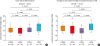
Table 2 presented the proportion of subjects who developed AD at 1 year according to subjects' cord-blood LTL. Shorter telomere length did not increase the proportion of AD in either group (odds ratio [OR], 0.833; 95% confidence interval [CI], 0.301-2.309; P = 0.799). Moreover, further analyses showed that shorter telomere length did not influence the odds of the AD in either high-stress or low-stress group (high-stress group: OR, 0.927; 95% CI, 0.184-4.687; P = 1.000; low-stress group: OR, 0.500; 95% CI, 0.119-2.100; P = 0.473).
Table 2
Proportion of atopic dermatitis development according to the level of cord blood telomere length in each stress group

DISCUSSION
Prenatally-stressed groups had already presented shorter LTL at birth than less-stressed ones. This difference remained significant until subjects became 1 year old. On the other hand, cord blood LTLs were not different between subjects who were doomed to develop the AD and who were not, neither were the ones of the 1-year peripheral blood. When we reclassify subjects according to subjects' prenatal stress and the cord-blood LTL, shorter cord-blood LTLs displayed no significant increase in the proportion of AD development in either high-stressed or low-stressed groups, which implicates the LTL shortening is not a risk factor for AD development up to the age of 1. This study is the first attempt to validate the oxidative stress markers for the increased risk for AD development.
The mean LTL measured in this study from all cord blood samples was longer than that of previous reports.1618 One study on the infant from mainly hypertensive mothers reported a mean cord blood LTL of 10.95 ± 0.09 kbp in males and 11.07 ± 0.08 kbp in females.16 Another study in the United States reported a mean cord blood LTL of 6.98 ± 0.41 kbp from the high-stressed groups and of 8.74 ± 0.24 kbp from the low-stressed ones, but their mothers consisted mainly of a specific ethnicity, African Americans.18 We believe this discrepancy would not doubt the accuracy of our LTL measurement because ethnicity can affect the LTL and no reference value is known for Korean babies.19 Moreover, we assessed the LTL by measuring the mean TRF length via Southern blots, which is more accurate than the quantitative polymerase chain reaction analysis.20 We assigned 1 dedicated person and 2 assistants in measuring the mean TRF length, which would minimize the interpersonal variation.
The 1-year peripheral blood LTLs as well as the cord blood ones were short in subjects who were exposed to high prenatal maternal stress. Although it has recently been replicated that prenatal maternal stress shortens the offspring's LTL at birth,2122 no information is available until when the prenatal stress may affect the LTL shortening significantly. This study shows the impact of prenatal stress exposure may persist for at least the first year of one's life significantly. Moreover, the impacts are comparable to that of AD development because 1-year LTL in the HSOD group presented the comparable shortening with those in the HSWD as well as the LSWD group and a significant shortening than those in the LSOD group.
The shorter cord blood LTL in the prenatal stress-exposure group was consistent with our previous report that the ratio of placental reduced GSH and GSSG was decreased following maternal prenatal stress exposure.11 Considering that both telomere shortening and the reduced ratio of GSH/GSSG are associated with oxidative stress exposure,68 prenatal stress may have exposed the fetus to more oxidative stress. Moreover, since oxidative stress is thought to be related to AD,5 these findings justify our hypothesis that LTL could play a role of the biomarker that reflects the exposure to prenatal stress and that predict the risk of AD development at the same time. Contrary to our assumption, even though the oxidative stress may be associated with AD, it seems not directly or specifically induce AD development because LTL shortening was not different between HSWD and HSOD groups, and the shortening was most prominent in the LSWD group during the first year of life. In other words, shortened LTL may not be interpreted to induce AD development for itself; instead, some AD-inducing factors other than prenatal maternal stress may also have affected LTL shortening during the first year of life.
In this study, we further evaluated the proportion of AD development in 4 groups of subjects that were reclassified according to the exposure to prenatal maternal stress and the LTL shortening, by which we aimed to assess the additive role of LTL shortening when it was combined with the exposure to prenatal maternal stress. As a result, the proportion of AD development was not increased by the shorter LTL compared with the others in either prenatally high-stressed or low-stressed groups. This result also discards the efficacy of LTL as a marker for later AD development.
We should mention some limitations of our study. First, we have confined AD to subjects that had AD before the age of 1 year, which may be complicated by subjects not-yet, but later showing AD. Nonetheless, we had to choose the age of 1 year because 60% of subjects with AD presents symptoms during the first year of life and, even in an Asian cohort, 78.5% of AD subjects were diagnosed before 12 months of age.2324 Moreover, before the age of 2 subjects begin to present their respiratory allergies or to outgrow their AD, which may complicate assessing the relationship between the LTL and AD development. Secondly, we have not assessed LTL from the whole population: the shortage of financial support led us to analyze samples of 4 typical groups of subjects instead, which eventually limits the findings to be generalized in the whole population. Thirdly, we could not present specific pathways that can replace the oxidative stress, by which the prenatal maternal stress would make subjects prone to AD development. Despite all these limitations, our study is still valuable to have investigated the relationship between the prenatal stress-exposure, LTL, and the AD development from the birth cohort samples.
In conclusion, cord blood LTL was shorter in prenatally stressed infants than in unstressed ones, which difference was still significant when subjects became 1 year old. Considering that cord blood LTL was not different according to later AD development at 1 year, shorter LTL made no increase in the proportion of later AD development in either prenatally high-stressed or low-stressed groups. These results implicate that the LTL shortening is not a risk factor for increasing AD development until the age of 1, and a longer investigation may be necessary for validation. Currently, the results doubt the role of LTL shortening as a marker for risk assessment tool for the maternal stress-associated AD development in the offspring.
 XML Download