

 PDF
PDF ePub
ePub Citation
 Print
Print


Abstract
A primary benign schwannoma of the liver is extremely rare. Only 30 cases have been reported in the medical literature worldwide, and only one case has been reported in Korea previously. A 56-year-old man was admitted to Gil Medical Center with incidental findings of a hepatic mass by abdominal computed tomography. The computed tomography and magnetic resonance image revealed a 3×2 cm-sized solid mass in the left lobe of the liver. Histological examination confirmed the diagnosis of a benign schwannoma, proven by positive immunoreaction with the neurogenic marker S-100 protein and a negative response to CD34, CD117, and smooth muscle actin. We report a primary benign schwannoma of the liver and review the literature.
References
1. Albert P, Patel J, Badawy K, et al. Peripheral nerve schwannoma: a review of varying clinical presentations and imaging findings. J Foot Ankle Surg. 2017; 56:632–637.

2. Yamamoto M, Hasegawa K, Arita J, et al. Primary hepatic schwannoma: a case report. Int J Surg Case Rep. 2016; 29:146–150.
3. Jung HI, Lee HU, Ahn TS, et al. Primary hepatic malignant peripheral nerve sheath tumor successfully treated with combination therapy: a case report and literature review. Ann Surg Treat Res. 2016; 91:327–331.
4. Yin SY, Zhai ZL, Ren KW, et al. Porta hepatic schwannoma: case report and a 30-year review of the literature yielding 15 cases. World J Surg Oncol. 2016; 14:103.
5. Momtahen AJ, Akduman EI, Balci NC, Fattahi R, Havlioglu N. Liver schwannoma: findings on MRI. Magn Reson Imaging. 2008; 26:1442–1445.
6. Young SJ. Primary malignant neurilemmoma (schwannoma) of the liver in a case of neurofibromatosis. J Pathol. 1975; 117:151–153.
7. Lee WH, Kim TH, You SS, et al. Benign schwannoma of the liver: a case report. J Korean Med Sci. 2008; 23:727–730.
8. Shih YC, Chen YL, Fang HY, Wu CY, Lin YC, Lin YM. Schwannoma mimicking liver tumor. Thorac Cardiovasc Surg. 2009; 57:436–439.
9. Ozkan EE, Guldur ME, Uzunkoy A. A case report of benign schwannoma of the liver. Intern Med. 2010; 49:1533–1536.
10. Madhusudhan KS, Srivastava DN, Dash NR, Gupta C, Gupta SD. Case report. Schwannoma of both intrahepatic and extrahepatic bile ducts: a rare case. Br J Radiol. 2009; 82:e212–e215.
11. Kulkarni N, Andrews SJ, Rao V, Rajagopal KV. Case report: benign porta hepatic schwannoma. Indian J Radiol Imaging. 2009; 19:213–215.
12. Yoshida M, Nakashima Y, Tanaka A, Mori K, Yamaoka Y. Benign schwannoma of the liver: a case report. Nihon Geka Hokan. 1994; 63:208–214.
13. Kim YC, Park MS. Primary hepatic schwannoma mimicking malignancy on fluorine-18 2-fluoro-2-deoxy-D-glucose positron emission tomography-computed tomography. Hepatology. 2010; 51:1080–1081.
14. Hayashi M, Takeshita A, Yamamoto K, Tanigawa N. Primary hepatic benign schwannoma. World J Gastrointest Surg. 2012; 4:73–78.
15. Ota Y, Aso K, Watanabe K, et al. Hepatic schwannoma: imaging findings on CT, MRI and contrastenhanced ultrasonography. World J Gastroenterol. 2012; 18:4967–4972.
16. Lederman SM, Martin EC, Laffey KT, Lefkowitch JH. Hepatic neurofibromatosis, malignant schwannoma, and angiosarcoma in von Recklinghausen's disease. Gastroenterology. 1987; 92:234–239.
17. Heffron TG, Coventry S, Bedendo F, Baker A. Resection of primary schwannoma of the liver not associated with neurofibromatosis. Arch Surg. 1993; 128:1396–1398.
18. Godlewski G, Sawan S, Targhetta P, Pignodel C, Marty-Double C, Gaujoux AF. A malignant schwannoma of the jejunum associated with multiple neurofibromas and a primary adenoma of the parathyroid. Ann Gastroenterol Hepatol (Paris). 1989; 25:13–17.
19. Ikehara H, Li Z, Watari J, et al. Histological diagnosis of gastric submucosal tumors: a pilot study of endoscopic ultrasonography-guided fine-needle aspiration biopsy vs mucosal cutting biopsy. World J Gastrointest Endosc. 2015; 7:1142–1149.
20. Harwalkar JA, Lee JH, Hughes G, Kinney SE, Golubić M. Immunoblotting analysis of schwannomin/merlin in human schwannomas. Am J Otol. 1998; 19:654–659.
Fig. 1.
Abdominal ultrasonography (US). US revealed an about 3 cm-sized, low echoic mass with homogeneous pattern in the left lobe of the liver.
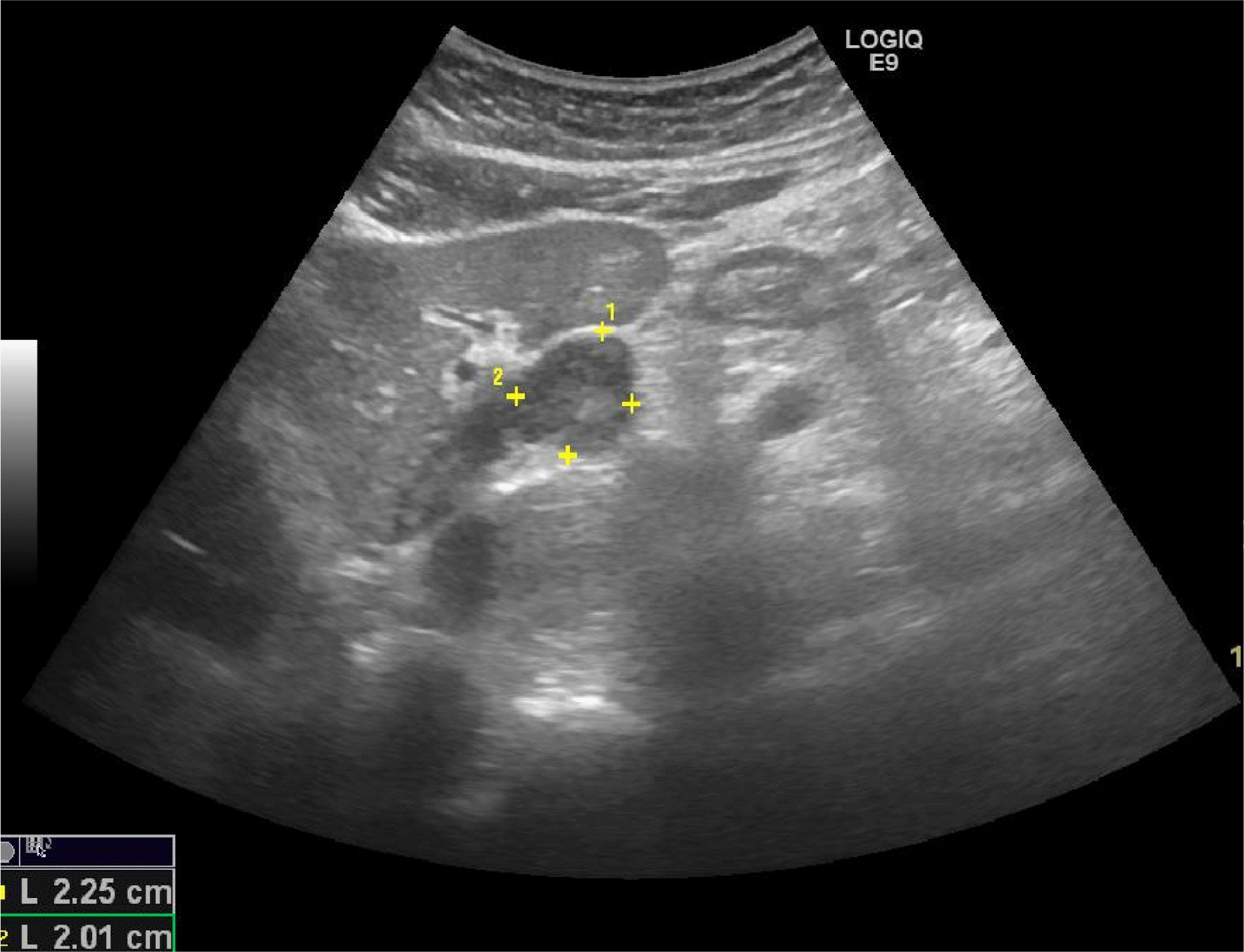
Fig. 2.
Dynamic abdominal computed tomography (CT). (A) Arterial phase (arrow), (B) portal phase (arrow), (C) venous phase: CT showed a well-defined, rounded (arrow), low-attenuating mass with homogeneous attenuation in the left lobe of the liver.
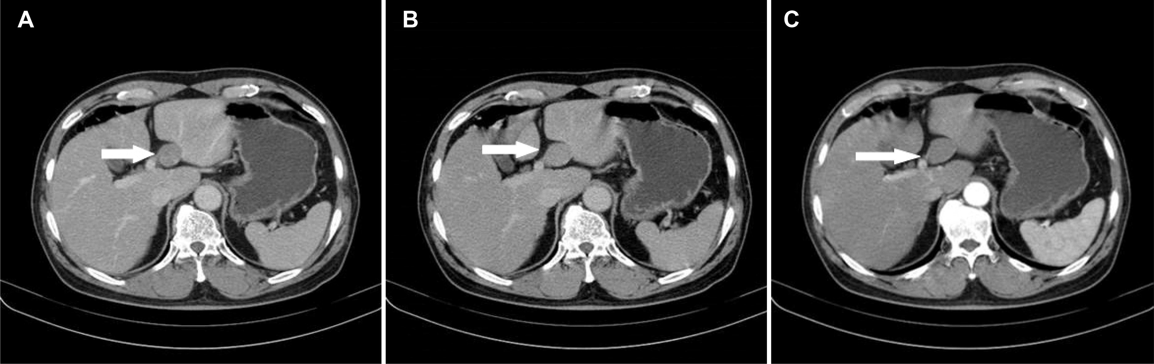
Fig. 3.
Liver magnetic resonance imaging. (A) T1-WI. A 35 mm suspicious enhancing lesion (white arrow) in the left liver: a mass that is mildly hypointense or isointense on non-contrasted T1-WI. (B) T2-WI. Lesion (white arrow) was heterogeneously hyperintense. (C) Hepatobiliary phase. Gadoxetic-acid enhanced heaptobiliary phase images show hypointense lesion (white arrow). WI, weighted image.
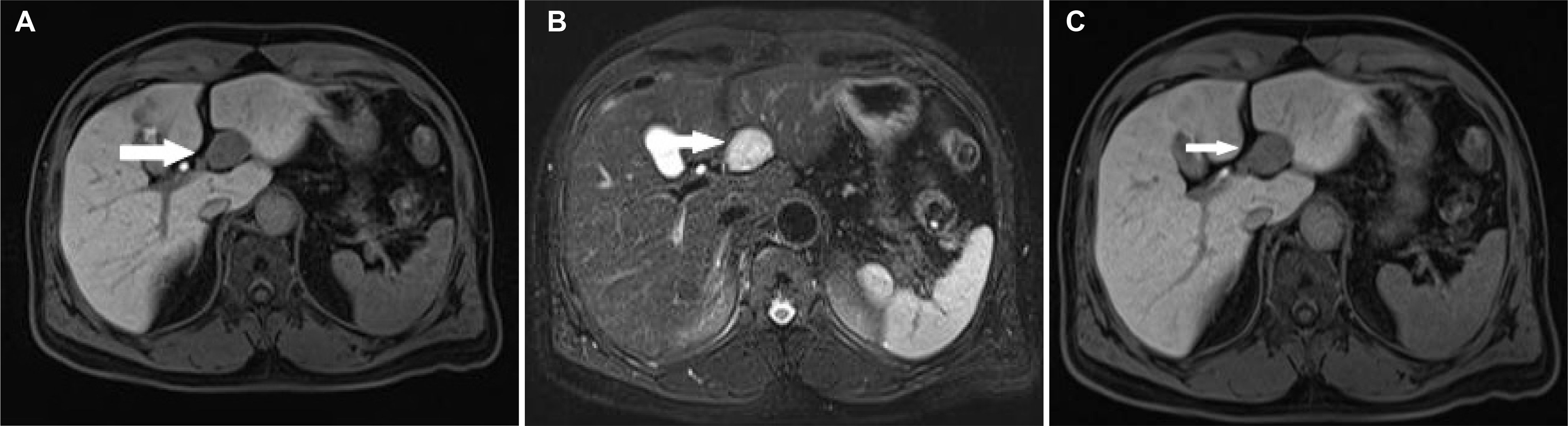
Fig. 4.
Histologic examination. (A) The sections show a moderately cellular schwannoma. Mitoses are inconspicuous (H&E, ×200). (B) The tumor is composed of bundles of compact spindle cells that are immunoreactive to S-100 protein, brown particles (immunohistochemical stain, ×200).
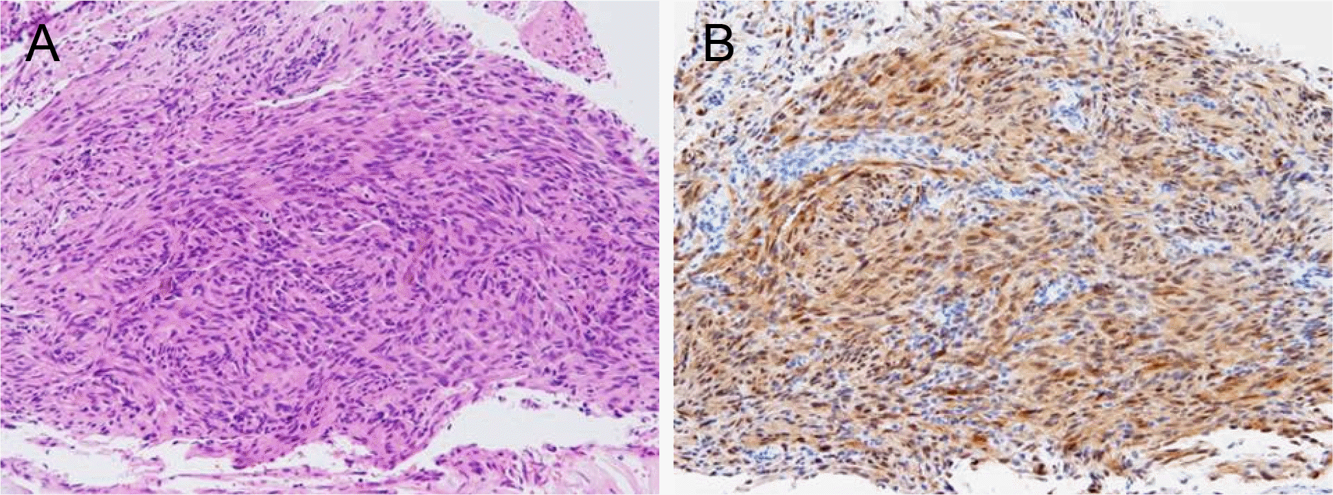
Table 1.
Summary of Clinicopathological Data from 30 Cases of Hepatic Schwannoma
 XML Download