

 PDF
PDF ePub
ePub Citation
 Print
Print


INTRODUCTION
Colonoscopy is a commonly performed endoscopic procedure to evaluate and treat various colorectal diseases.1 Although it is generally considered to be a safe procedure, it does have a risk of a potentially life-threatening complication, colorectal perforation.2
Colorectal perforation usually appears as pneumoperitoneum, if it occurs in the intraperitoneal portion of the organ. Rarely, however, colorectal perforation may cause an air leak into the retroperitoneal space, causing pneumoretroperitoneum if the perforation site is located in the regions attached to the extraperitoneal space, such as the posterior walls of the sigmoid, rectosigmoid, rectum, ascending, or descending colon.3 Pneumoretroperitoneum can further progress to pneumomediastinum, pneumothorax, and subcutaneous emphysema, especially if there is a large amount of air leakage through the perforation site.4
When colorectal perforation occurs, the patient typically complains of abdominal pain as an initial symptom. However, in case of extraperitoneal perforation, atypical symptoms, such as subcutaneous swelling with crepitus, chest discomfort, or dyspnea may develop.
We report a patient presenting a change in voice and neck swelling as chief complaints after diagnostic colonoscopy and esophagogastroduodenoscopy. The patient was diagnosed with a rectosigmoid perforation that led to pneumoretroperitoneum, pneumomediastinum, pneumothorax, and subcutaneous emphysema. The patient was successfully managed with non-surgical treatment.
CASE REPORT
A 64-year-old woman was transferred to our emergency department from a private clinic with chief complaints of a change in voice and neck swelling that developed after diagnostic colonoscopy and esophagogastroduodenoscopy, which were performed two hours prior to the onset of symptoms. Other symptoms included mild chest discomfort. The patient had a medical history of hypertension and diabetes mellitus.
The initial vital signs were blood pressure of 176/107 mmHg, a pulse rate of 76/min, a respiration rate of 22/min, and a body temperature of 37.8℃. The patient did not have a febrile sensation. With regard to the initial lab findings, her complete blood count was normal (white blood cell count 5,350/mm3, hemoglobin 14.0 g/dL, platelets 322,000/mm3), blood urea nitrogen and creatinine were normal (17.5/0.69 mg/dL), and blood C-reactive protein was normal (0.29 mg/dL). The initial arterial blood gas analysis revealed a slight hypoxia (pH 7.48, pCO2 34 mmHg, pO2 68 mmHg, HCO2 25.3 mmHg, SpO2 95%). Other lab findings were within normal ranges. No peritoneal irritation or respiratory distress signs were found upon physical examination. Crepitus was palpated around the neck.
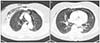
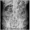
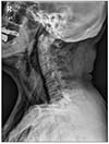
A posteroanterior chest x-ray and contrast-enhanced computed tomography (CT) of the chest showed large and diffuse subcutaneous emphysema around the neck and thorax wall, as well as pneumoretroperitoneum, pneumomediastinum, pulmonary interstitial emphysema, and pneumothorax (Fig. 1). An erect and supine abdominal x-rays also revealed pneumoretroperitoneum (Fig. 2). Anteroposterior and both lateral neck x-rays revealed prevertebral air and extensive subcutaneous emphysema around the neck (Fig. 3). Considering the distribution of free air, perforation of the extraperitoneal portion of the gastrointestinal organ attached to the retroperitoneal cavity was suspected.
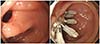
To rule out any possibilities of esophagus perforation, esophagography was performed. There was no evidence of contrast media leakage from the esophagus into the thoracic cavity. A colonoscopy was performed, with the quality of bowel preparation being fair. A visible perforation was found at the rectosigmoid colon (Fig. 4A). Endoscopic clipping was performed, and the perforation site was closed successfully with seven endoscopic clips (Fig. 4B).
After successful primary closure, we decided to manage the patient conservatively because the patient showed stable vital signs and was in good general condition without signs of peritoneal irritation. The patient was treated with bowel rest, intravenous nutrition support, systemic antibiotics, oxygen, and symptomatic care. A closed thoracotomy was considered due to pneumothorax, but was not performed since the amount of air in the thorax was relatively small and she did not suffer from any symptoms of respiratory distress.
Over the course of conservative care, symptoms improved, and the amount of free air gradually decreased according to simple x-rays. By the 8th day in hospital, abdominopelvic CT showed that pneumoretroperitoneum and pneumomediastinum were markedly decreased since the last CT scan, while pneumothorax was eliminated. The patient's voice also improved, and neck swelling and fever also subsided. On the 10th day in hospital, free air was no longer visible on simple x-rays. Oral intake was initiated and did not cause any problems. The patient was then discharged with oral antibiotics. The patient was later followed-up at our outpatient clinic and was confirmed to be fully recovered without any further complications.
Discussion
Colorectal perforation is a serious complication of colonoscopy and can result in extended hospital stays, operations, peritonitis, sepsis, multiple organ failure, and even death.5 A retrospective review of cases between 1980 and 2006 showed that when iatrogenic colonic perforations were managed operatively, the morbidity and mortality rates were 35% and 7%, respectively.6 Other studies have reported that the mortality rate can be as high as 25%.7 The rate of perforation during colonoscopy has been reported to be 0.1-0.3%.8 Therapeutic colonoscopy is known to have an increased risk of perforation compared with diagnostic-only colonoscopy. The perforation rate has been reported to be about 0.16% for diagnostic colonoscopies and about 0.44% for therapeutic colonoscopies.9 However, a review conducted by Iqbal et al. argued that a diagnostic colonoscopy does not necessarily carry a lower rate of perforation than therapeutic colonoscopy.6
Perforations can be intraperitoneal, extraperitoneal, or a combination of both, depending on the location of the perforation site; however, it has been shown that intraperitoneal perforations are much more common.10 In the lower gastrointestinal tract, extraperitoneal organs include ascending and descending colon, posterior walls of the sigmoid colon, and rectum.3 The sigmoid colon, including rectosigmoid, is known as the most frequent site of all types of colorectal perforation, followed by the cecum.6
The risk factors of iatrogenic colonic perforation are elderly patients, diverticulosis, severe colitis, inflammatory bowel disease, malignancy, pelvic adhesions due to history of abdominal or pelvic surgery, radiation therapy or inflammation, and lack of experience of the physician.11121314 The patient in our case had none of the above risk factors.
In our case, perforation caused an intraluminal air leak that accumulated in the retroperitoneal space. Since the retroperitoneum, mediastinum and thorax are anatomically connected, extraluminal free air reached various compartments of the body and caused pneumomediastinum, pneumothorax and subcutaneous emphysema.1516 Free air in one of these spaces can travel to nearby structures, including the fascial planes and large vessels.17 Large amount of air may rupture the mediastinal pleura, penetrating the pleural cavity.18 Alternatively, any free air in the peritoneal cavity can permeate through small diaphragmatic fenestrations and enter the pleural space.18 Pneumothorax resulting from colonic perforations is very rare. Indeed, a review by Zeno et al. found only nine cases of pneumothorax; of which only two occurred after diagnostic colonoscopy.11
Patients with perforations most commonly suffer from abdominal pain. Fever with leukocytosis and tachycardia may also develop. Physical examination may reveal a rigid abdomen with tenderness, rebound tenderness, and muscle guarding. However, in extraperitoneal perforation cases, abdominal pain may not be the initial symptom. Tiwari et el. conducted a review of 32 extraperitoneal perforation cases and concluded that subcutaneous emphysema of the neck, face, or upper chest was the most common clinical finding, with a prevalence rate of 65%.17 Palpable crepitus is usually accompanied with subcutaneous emphysema. Abdominal pain, on the other hand, was seen in only 34%, and dyspnea was seen in 25%.17 Nearly 10% of patients remained asymptomatic.17 Therefore, even if there is no abdominal pain, perforation should be considered if atypical symptoms occur after a colonoscopy. Rare but possible atypical complications include pneumopericardium, periorbital swelling, pharyngeal swelling, and pneumoscrotum.1920
Symptoms of perforation may appear after several hours.20 A review conducted by Tiwari et al. revealed that 52% of perforations were detected immediately or within 1 hour, whereas 29% were found within 1-24 hours and 19% found after 24 hours from the procedure.17 In our case, perforation was not detected immediately when it first happened at the private clinic because the patient did not show any symptoms during and immediately following the colonoscopy. Therefore, upon completion of the colonoscopy, esophagogastroduodenoscopy was performed next as planned. After the esophagogastroduodenoscopy, neck swelling was then observed as the initial symptom, accompanied with a change in voice and mild chest discomfort. Because of these unusual symptoms with a delayed onset, the patient was transferred to our hospital for further evaluation.
Endoscopic closure was done successfully in our case because the perforation size remained relatively small, as the patient was transferred to our hospital in a timely manner. In addition, bowel preparation was done properly so that fecal materials did not contaminate the peritoneal space. Furthermore, systemic antibiotics were quickly administered. All of these factors contributed to a successful treatment of perforation without the need for surgery.
In conclusion, we presented a rare case of rectosigmoid perforation occurring after a diagnostic colonoscopy that led to the extensive amount of extraperitoneal free air development with voice change and neck swelling as the initial symptoms. Atypical symptoms should not be overlooked by physicians as they can be indicative of perforation.
 XML Download