

 PDF
PDF ePub
ePub Citation
 Print
Print


INTRODUCTION
Gastric adenoma is usually classified using a two-grade system: low-grade dysplasia (LGD) and high-grade dysplasia (HGD).123 The clinical importance of gastric adenoma has been emphasized due to its close association with the risk of developing gastric cancer.2 Gastric adenoma is generally accepted as a precancerous lesion. In most instances, it remains stable or regressed, although it can progress at a slow rate.1 Thus, it can be removed by an endoscopic resection or surgical resection, when possible.4 In particular, endoscopic submucosal dissection (ESD) is not limited by resection size and is expected to replace surgical resection. However, it is still associated with a higher incidence of complications than standard endoscopic mucosal resection (EMR) procedures, requiring a high level of endoscopic skill.56 Considering its limitation, new feasible treatment methods are required for gastric adenoma. Argon plasma coagulation (APC) is a method of contact-free electrocoagulation, whereby energy is transmitted to the tissue through an ionized argon gas.78 APC has widely been used to treat many gastrointestinal diseases, including bleeding peptic ulcer, watermelon stomach, hemorrhagic proctitis, and esophageal varices.910 Recently, its application has been broadened into the field of treatment for gastric adenoma.11 The purpose of this study was to investigate the clinical efficacy and to assess the usefulness of APC in treating gastric adenoma compared with ESD.
SUBJECTS AND METHODS
1. Patients
We retrospectively reviewed patients with gastric adenoma, who underwent treatment endoscopically at Inje University Ilsan Paik Hospital, between January 2006 and June 2013. All lesions had been identified with endoscopic examination at our institution. Before APC or ESD treatment, endoscopic examination was performed, and at least three biopsy specimens were taken from the lesions. Treatment methods, classified as either APC or ESD, were chosen at the discretion of each oeprating physician. Three endoscopists performed both diagnostic and therapeutic procedures. Each endoscopic report was reviewed to identify the lesion size and gross appearance. After discharge, follow-up endoscopic examinations with biopsy were performed at 3, 6, and 12 months post-operation, then every 12 months thereafter. Patients who were previously treated at another hospital or those who were lost to follow-up were excluded from the final analysis: Five patients had undergone prior treatment at another hospital, and nine had failed to successfully be followed-up for more than three months. The largest lesion was selected in the analysis if a patient had multiple adenoma. Finally, a total of 210 patients with gastric adenomas were included for analysis. Patients were treated with either APC (97 patients) or ESD (113 patients).
2. Endoscopic procedures and complications
All lesions were removed by ESD or APC. Both treatments were performed under sedation with midazolam. The APC group received APC ablation (VIO 300D with APC 2; Erbe Elektromedizin GmbH, Tuebingen, Germany; argon gas flow rate was 1.8 L/min, with pulsed argon plasma coagulation, 40 Watt [W]) after submucosal saline injections to prevent procedure-related complications. In the ESD group, injections of submucosal saline, circumferential incision, and submucosal dissection were performed by needle knife and IT knife (Olympus, Tokyo, Japan). A complete resection was defined as one in which the resected tumor had tumor-free lateral and deep margins. Tumor recurrence was confirmed histologically, using biopsy specimens obtained from the initially treated lesions during the follow-up period. With respect to complications, bleeding was defined as a decrease in blood hemoglobin level of more than 2 g/dL, accompanied by the occurrence of hematemesis, melena, or the combination of unstable vital signs with fresh blood or clots upon Levin tube irrigation within the first 4 weeks after endoscopic treatment. Perforation was classified as macro- and microperforation. Macroperforation was readily recognized endoscopically, while microperforation was detected by the presence of free air on plain radiographs taken after the procedure.
3. Clinical analysis
Clinical, pathologic data, treatment modalities, and outcomes were collected from the medical records of all patients. Clinical data included sex, age, tumor site, tumor size, and macroscopic morphology. The types of macroscopic morphology were classified as elevated, flat, or depressed, as proposed by the Paris classification.12 We compared the clinical and pathologic characteristics, admission rates, mean duration of hospital stay, complications, and recurrence rates between the two groups. The study protocol was approved by the institutional review board of Ilsan Paik Hospital, Inje University College of Medicine, Goyang, Korea.
4. Statistical analysis
To analyze the baseline characteristics, Pearson's chi-squared and Fisher's exact tests were used for categorical variables and t-test was used for continuous variables. Continuous variables are shown as the mean and standard deviation. Statistical significance was set at a p-value of less than 0.05. We performed a subgroup analysis among APC patients by the admission status (i.e., APC-inpatient basis vs. APC-outpatient basis). The statistical analysis was carried out using SPSS version 17.0 software (SPSS INC., Chicago, IL, USA).
RESULTS
1. Patients and baseline clinical characteristics
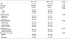
A total of 210 patients were eligible for this study: 97 patients were treated with APC and 113 patients with ESD. The baseline characteristics of the 210 patients are summarized in Table 1. The mean age of patients was 65.8 years for APC and 64.6 years for ESD (p=0.136). There were larger tumors in the ESD group than in the APC group (1.1±0.7 cm vs. 0.9±0.4 cm, p<0.001). There were more lesions on the upper or middle third locations in vertical locations in the APC group, but lower third locations were more frequent in the ESD group (p=0.008). Moreover, there were more lesions in the greater curvature on the horizontal locations in the ESD group (p=0.01).
The proportion of high-grade dysplasia was 2.1% and 23.9% in the APC group and ESD group, respectively (p<0.001). There were more adenomas with elevated morphology in the ESD group than in the APC group (79.6% vs. 60.8% p=0.01).
2. Local recurrence, complication, admission, and cost comparison between the APC and ESD group
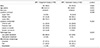
Following the procedures, 97 patients in the APC group and 113 patients in the ESD group had regularly follow-up visits. The median follow-up duration for the APC and ESD groups was 6.0 (range 3-36) months and 12.0 (range 3-48) months, respectively. En bloc resection was attempted in all ESD cases, with a complete resection rate of 89.4 % (101 of 113). In 11 patients, the lateral margin of the resected specimen was histologically positive. The residual tumor was treated by APC, and there was no recurrence during the follow-up period after APC. In one patient, the vertical margin of the resected specimen was histologically determined to be early gastric cancer, and surgery was carried out. There was no recurrence during the follow-up period after surgery. Recurrence rates were higher in the APC group than in the ESD group (15.3% vs. 3.5%, p=0.003) (Table 2).
Complications did not occur in the APC group. However, one case of microperforation (0.9%) and six cases of bleeding (5.3%) occurred in the ESD group (Table 2). The six patients who had postprocedural bleeding were successfully managed using an endoscopic hemostatic technique (hemoclip or electric coagulation). No patient required surgery or angiographic intervention. The admission rate was significantly higher in the ESD group than in the APC group (100% vs. 43.3%, p<0.001). The mean duration of hospital stay was significantly shorter in the APC group than in the ESD group (1.6 days vs. 5.8 days, p<0.001). The medical cost was significantly higher in the ESD group than in the APC group (1,430,610 won vs. 377,172 won, p<0.001) (Table 2).
3. Subgroup analysis among APC cases
The subgroup analysis by admission status was performed among patients who were treated by APC. Table 3 shows clinical characteristics between the two groups stratified by admission status. The subgroup analysis included 97 patients. Among those treated by APC, 55 patients (56.7%) were outpatients and 42 patients (43.3%) were inpatient. There were no significant differences with respect to the characteristics between two groups, except vertical location.
Table 4 shows the treatment results between the two groups. There was no significant difference between the two groups in the recurrence rate (APC-outpatient 16.4% vs. APC-inpatient 14.3%). No complications were observed in the APC group, on an outpatient or inpatient basis. However, medical costs were significantly lower in the APC outpatient group than in the APC inpatient group (292,842 won vs. 1,079,698 won, p<0.001).
4. Clinical characteristics, endoscopic findings, and treatment sequence of patients with local recurrence for APC
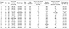
The local recurrence of the APC group was frequently observed as the elevated type (14 of 15) (Table 5). Otherwise, of all the flat lesions, no recurrence was observed (0 of 33). Four patients with adenomas ≥2.0 cm were treated with APC, and 2 of them experienced recurrence. There was a significant difference in the recurrence rate between adenomas ≥2 cm (50%, 2/4) and adenomas <2 cm (13.9%, 13/93) (p=0.047). However, there was no statistical difference in the recurrence rate between adenomas <1 cm (10.7%, 6/56) and adenomas 1-2 cm (18.9%, 7/37) (p=0.253). There was no difference in the recurrence rate according to vertical location (p=0.504). All patients (except one, who was lost to follow-up) were treated using APC or ESD or surgery. Six patients were treated by repeat APC. Seven patients were retreated by ESD. One patient was treated by surgery because the pathology was noted as early gastric cancer type IIa. There was no recurrence in six patients after repeat APC (Table 5).
DISCUSSION
Gastric adenoma is generally accepted as a precancerous lesion, and neoplastic risk differs by the type of dysplasia.1 LGD is known to regress in 38-49% of patients, while persist in 19-28%, and progress to HGD in 1-15%. HGD is known to regress in 5% of patients, persist in 14%, and progress in 81-85%.2 Although most cases of LGD are believed to regress or persist, some cases have been reported to progress to HGD or carcinoma after the median follow-up period, ranging from 34.5 to 41.5 months in one study.13 For this reason, endoscopic resection methods, such as EMR and ESD, are already recognized as the standard treatments of premalignant lesions and small intramucosal cancerous lesions.14 Although APC has been proven to be efficient for the treatment of mucosal and even submucosal cancers, it is typically applicable in only limited cases for patients who are not suitable for surgical resection or EMR/ESD due to the difficulty in predicting the depth of invasion and the inability to perform pathological evaluations, which are possible with complete resection.1516 Therefore, either surgical or endoscopic resection is usually preferred to APC treatment for patients whose physical conditions are strong enough to tolerate the resection procedures. The biological behavior of adenomas is quite different from that of carcinomas. Because adenomas are limited and confined to the mucosal layer, gastric adenomas with LGD have no capacity to invade or infiltrate into the deeper layers of the gastric wall.117 As APC at an optimal setting can totally cauterize the mucosal layer and superficial portion of the submucosal layer, in theory, gastric adenomas can be eradicated with APC. Different from gastric adenomas with HGD, which should be treated as early gastric cancers due to the risk of synchronous carcinoma and high risk of malignant progression, gastric adenomas with LGD rarely contain carcinomatous lesions at the time of diagnosis.1819 Based on these characteristics, APC treatment has many merits over endoscopic resection and frequent endoscopic follow-up, including short procedure time, low risk of bleeding and perforation, low procedure cost, and no requirement of hospitalization.11 Therefore, APC can be regarded as a reasonable treatment method for gastric adenomas with LGD.
Previous reports have indicated a local recurrence rate of 4-10% after APC in cases of early gastric cancer or gastric adenoma.15162021 However, to the best of our knowledge, an analysis of risk factors for local recurrence has not yet been performed. Our present study showed a high rate of local recurrence (15.3%) compared with other studies. The local recurrence of treated adenomas using APC in our study was frequently observed as the elevated type (14 of 15). Otherwise, no recurrence was observed in all the flat lesions (0 of 33). Moreover, the local recurrence of treated adenomas using APC was frequently observed to be large. There was no statistical significance; however, adenomas with a size of 1-2 cm tended to have a higher recurrence rate than those <1 cm (18.9% vs. 10.7%, p=0.253). Four patients with adenomas ≥2.0 cm were treated with APC, and two of them showed recurrence. This was potentially due to the insufficient coagulation depth that may occur from using the APC therapy, since the tumor volume is greater in the elevated lesions than in the flat type lesions of the same size. Therefore, APC may be a satisfactory and effective modality for flat lesions with a relatively small size. This finding has also been reported in other studies.1121
Other studies observed that all recurrent lesions following APC were superficial elevated type.1121 Moreover, a recent study showed that non-lifting after submucosal saline injection and a lower power setting of 40 W are both risk factors for local recurrence after APC, with a lower reported rate of local recurrence in lifting and 60 W or 80 W groups than in non-lifting and 40 W groups.22 In that study, the authors concluded that a higher power setting of 60 W or 80 W may be safe and effective for treating gastric neoplasms by APC.22 To reduce the recurrence rate, additional sessions of APC and changing the power setting in accordance with the tumor volume may be necessary. In addition, the depth of affecting tissue will likely increase as the APC application time over the same area increases. Therefore, the physician should treat with an activation time that corresponds with the desired effect.
In this study, among the 15 patients with recurrent lesions treated with an APC procedure, 6 patients were treated by secondary APC. There was no recurrence in all six repeated APC cases. Thus, even if recurrence is more frequent following APC than ESD, repeated APC treatments can be a good option for local recurrence. Certainly, regular follow-up is needed after APC to monitor for any local recurrence.
With respect to complications, perforation occurred in one patient who underwent ESD. However, there were no perforations in the APC group. APC-related perforation is very rare, accounting for about 0.3% in one study.23 This is much lower than with ESD (1-6%).242526 The perforation was caused by excessive tissue coagulation. Therefore, careful treatment with APC does not cause as much of a perforation compared with ESD. We did not find any procedure-related bleeding during and immediately after APC. However, in six cases (5.3%) after ESD, there was an occurrence of bleeding.
In our study, APC showed to be more advantageous for gastric adenoma compared with ESD, with respect to low admission rate, shorter duration of hospital stay, and low medical costs. The mean admission rate in the APC and ESD groups was 43.3% and 100.0%, respectively. The mean duration of hospital stay in the APC and ESD groups was 1.6 days and 5.8 days, respectively. The duration of hospitalization for ESD patients was longer than usual (2-3 days). In recent years, hospitalization period for ESD is relatively short, but the hospitalization period for ESD in our hospital was relatively long to observe complications at the beginning of ESD. The mean medical costs for APC outpatient, APC inpatient, and ESD groups were 292,842 won, 1,079,698 won, and 1,430,610 won, respectively. Moreover, it is noteworthy that there was no significant difference regarding the recurrence rate in the APC groups, on an outpatient and admission basis. In addition, no complication was observed in both groups. This suggests that APC is an effective and safe treatment modality for gastric adenoma on an outpatient basis. The APC group on an outpatient basis was shown to be five times more economical than ESD. Actually, some patients preferred APC to ESD due to hospital admission issues or economic considerations. Endoscopic resection remains technically difficult and cannot be performed in some cases due to high risk of bleeding or perforation as well as due to non-lifting after submucosal saline injection. APC may be one alternative treatment that is less invasive for patients with high risk of ESD-related complications. Moreover, APC does not differ according to endoscopic experience. It shows equal therapeutic outcomes between experienced and non-experienced endoscopists. Therefore, non-experienced endoscopists could treat small gastric adenomas that do not require a long training period, such as that required for ESD.1121
There are several limitations to this study. First, this was a single-center study with a limited number of patients. Second, the retrospective nature of this study may have resulted in a selection bias, which may have likely played a role in the choice of treatment modality (APC vs. ESD). In this study, there were more adenomas with larger size and with elevated type in the ESD group than in the APC group. There were more lesions on the lower-third location in the ESD group. Moreover, the proportion of high-grade dysplasia was higher in the ESD group than in the APC group. In addition, short follow-up duration may be another limitation. Further randomized prospective studies are necessary to confirm that APC is a more effective and reasonable treatment for gastric adenomas compared with ESD.
In conclusion, this study demonstrated that gastric adenoma can be safely treated by APC without serious complications. Due to its association with minimal hospital admission, low medical cost, and favorable outcomes, as well as very low complication rates, APC may be a good treatment option, especially for relatively small, flat adenomas. However, regular endoscopic follow-up is necessary to detect any residual or recurrent lesions, as it has been shown to have a relatively higher rate of local recurrence after APC.


 XML Download