References
1. Bernstein CN, Eliakim A, Fedail S, et al. World gastroenterology organisation global guidelines inflammatory bowel disease: update August 2015. J Clin Gastroenterol. 2016; 50:803–818.
2. Yang SK, Yun S, Kim JH, et al. Epidemiology of inflammatory bowel disease in the Songpa-Kangdong district, Seoul, Korea, 1986–2005: a KASID study. Inflamm Bowel Dis. 2008; 14:542–549.

4. Kim YS, Kim YH, Lee KM, Kim JS, Park YS. IBD Study Group of the Korean Association of the Study of Intestinal Diseases. Diagnostic guideline of intestinal tuberculosis. Korean J Gastroenterol. 2009; 53:177–186.
5. Moran CP, Neary B, Doherty GA. Endoscopic evaluation in diagnosis and management of inflammatory bowel disease. World J Gastrointest Endosc. 2016; 8:723–732.

6. Lee JM, Lee KM. Endoscopic diagnosis and differentiation of inflammatory bowel disease. Clin Endosc. 2016; 49:370–375.

7. Park JB, Yang SK, Myung SJ, et al. Clinical characteristics at diagnosis and course of Korean patients with Crohn's disease. Korean J Gastroenterol. 2004; 43:8–17.
8. So H, Ye BD, Park YS, et al. Gastric lesions in patients with Crohn's disease in Korea: a multicenter study. Intest Res. 2016; 14:60–68.

9. Ye BD, Jang BI, Jeen YT, et al. Diagnostic guideline of Crohn's disease. Korean J Gastroenterol. 2009; 53:161–176.
10. Turner D, Griffiths AM. Esophageal, gastric, and duodenal manifestations of IBD and the role of upper endoscopy in IBD diagnosis. Curr Gastroenterol Rep. 2009; 11:234–237.

11. Kang MS, Park DI, Park JH, et al. Bamboo joint-like appearance of stomach in Korean patients with Crohn's disease. Korean J Gastroenterol. 2006; 48:395–400.
12. Fujiya M, Sakatani A, Dokoshi T, et al. A Bamboo Joint-Like Appearance is a Characteristic Finding in the Upper Gastrointestinal Tract of Crohn's Disease Patients: A Case-Control Study. Medicine (Baltimore). 2015; 94:e1500.
Fig. 1.
Colonoscopic finding. (A) In the terminal ileum, diffuse hyperemic mucosa with a few tiny erosions was noted. (B) In the whole colon, multiple longitudinal, deep active ulcers with normal looking intervening mucosa were scattered.

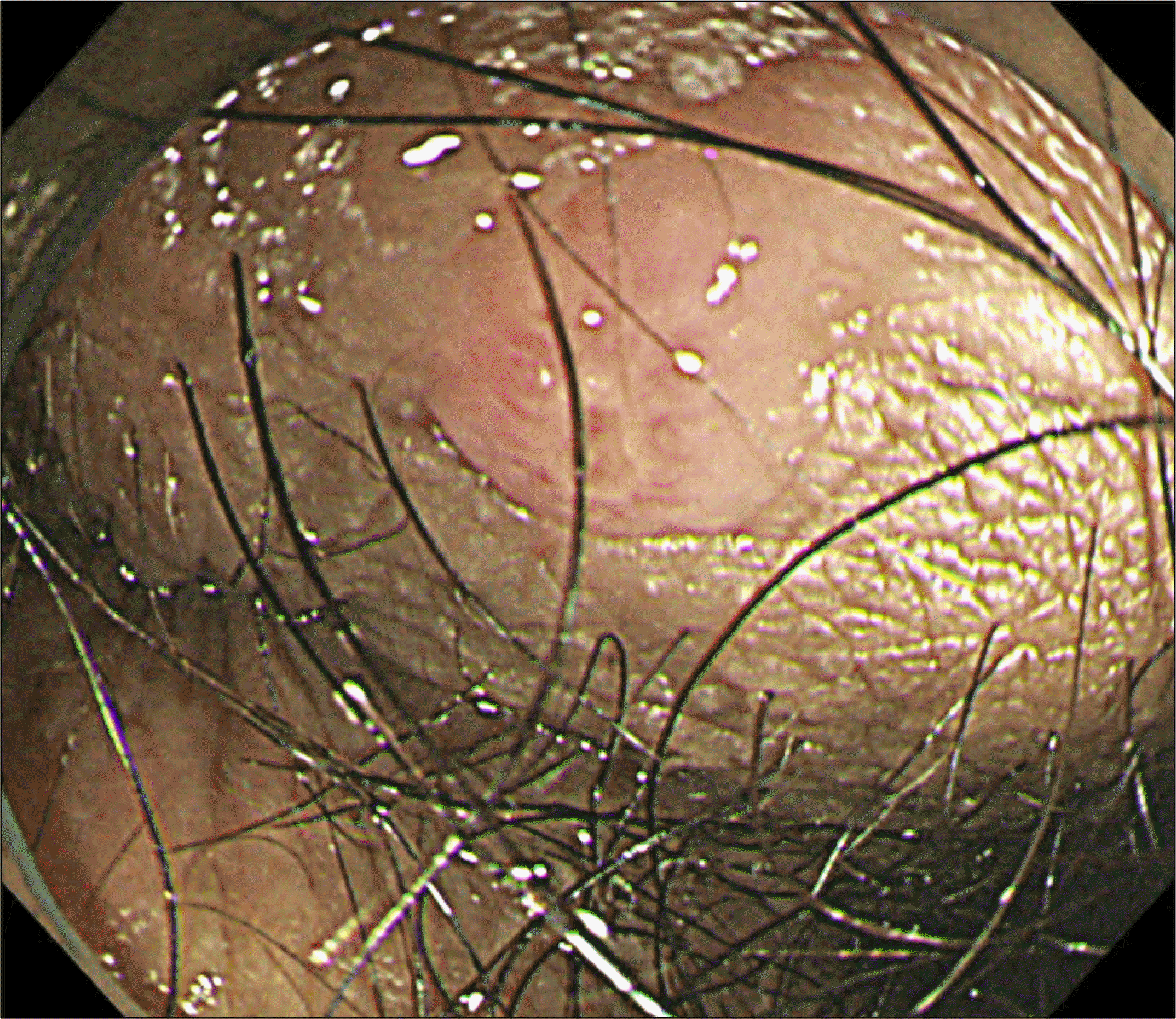
Fig. 2.
Perianal finding with a colonoscopy. A raised 4 mm-sized erosion― round in shape― was noted in the perianal skin.


 PDF
PDF ePub
ePub Citation
 Print
Print


 XML Download