

 PDF
PDF ePub
ePub Citation
 Print
Print


Abstract
Background/Aims
Several studies suggest that pyogenic liver abscess (PLA) is associated with colon neoplasm. A colonoscopic exam for cryptogenic PLA might detect a hidden colon neoplasm, through which intestinal flora can be transmitted into the liver. However, there are no prospectively enrolled cross-sectional data for colonic neoplasm in cryptogenic PLA.
Methods
Patients with PLA were prospectively enrolled from two university hospitals. Among them, all the patients with cryptogenic PLA were recommended for colonoscopic exam to check for colonic neoplasm.
Results
One hundred eighty-three patients with PLA were enrolled in the study for 22 months. One hundred and one (55.2%) patients did not have a definite cause of liver abscess at initial evaluation. The median diameter of the largest lesion was 5.7 cm (1.0–14.0 cm), and 74.3% of the patients were treated by percutaneous abscess drainage. Ninety-one percent of the patients who had an identified pathogen yielded Klebsiella. Sixty-two patients underwent colonoscopic exams, and no one had a colonic cancer, one had an adenomatous polyp with high grade dysplasia (1.6%), and 27 had adenomatous polyps with low grade dysplasia (43.5%; 41.0% in male and 43.5% in female). Of fifty patients who underwent an esophagogastroduodenoscopic exam, nine had gastric ulcers, one had an esophageal ulcer, and one had hemorrhagic gastritis.
References
1. Jeong SW, Jang JY, Lee TH, et al. Cryptogenic pyogenic liver abscess as the herald of colon cancer. J Gastroenterol Hepatol. 2012; 27:248–255.

2. Koo HC, Kim YS, Kim SG, et al. Should colonoscopy be performed in patients with cryptogenic liver abscess? Clin Res Hepatol Gastroenterol. 2013; 37:86–92.
3. Lai HC, Lin HC. Cryptogenic pyogenic liver abscess as a sign of colorectal cancer: a population-based 5-year follow-up study. Liver Int. 2010; 30:1387–1393.
4. Kim AY, Chung RT. Bacterial, parasitic, and fungal infections of the liver, including liver abscess. Feldman M, Friedman LS, Brandt LJ, editors. Sleisenger and Fordtran's gastrointestinal and liver disease. 10th ed.Philadelphia, PA: Saunders;2016. p. 1374–1392.
5. Antia FP, Marker F. Hepatic abscess secondary to duodenal ulcer. Lancet. 1955; 268:649–650.
6. Heo NY, Park SH, Park J, et al. Pyogenic liver abscess with complicating intestinal tuberculosis. J Med Cases. 2013; 3:370–372.
7. Knowels R, Rinaldo JA. Pyogenic liver abscess probably secondary to sigmoid diverticulitis: report of two cases. Gastroenterology. 1960; 38:262–266.
8. Margalit M, Elinav H, Ilan Y, Shalit M. Liver abscess in inflammatory bowel disease: report of two cases and review of the literature. J Gastroenterol Hepatol. 2004; 19:1338–1342.
9. Song J, Swekla M, Colorado P, Reddy R, Hoffmann S, Fine S. Liver abscess and diarrhea as initial manifestations of ulcerative colitis: case report and review of the literature. Dig Dis Sci. 2003; 48:417–421.
10. Cancer incidence according to the age group. [Internet]. Goyang: National Cancer Information Center; [updated 2015 Dec 23;cit-ed 2016 Jul 17]. Available from:. http://www.cancer.go.kr/mbs/cancer/subview.jsp?id=cancer_040103000000.
11. Lai HC, Lin CC, Cheng KS, et al. Increased incidence of gastrointestinal cancers among patients with pyogenic liver abscess: a population-based cohort study. Gastroenterology. 2014; 146:129–137.e1.
12. Yang MH, Rampal S, Sung J, et al. The prevalence of colorectal adenomas in asymptomatic Korean men and women. Cancer Epidemiol Biomarkers Prev. 2014; 23:499–507.
13. Jung HG, Kim DH, Lee CH. A case of subcapsular liver abscess secondary to perforating ulcer of gastric cancer. Korean J Gastroenterol. 2010; 56:109–113.
14. Okuno M, Adachi S, Nakamura N, et al. A case of advanced gastric cancer with liver abscesses. Nihon Shokakibyo Gakkai Zasshi. 2013; 110:869–874.
15. Sakata T, Narita M, Ohtani N, et al. A case of phlegmonous gastritis with multiple liver and splenic abscesses. Nihon Shokakibyo Gakkai Zasshi. 2011; 108:50–58.
16. Lin JN, Lin CL, Lin MC, Lai CH, Lin HH, Kao CH. Pyogenic liver abscess in patients with inflammatory bowel disease: a nationwide cohort study. Liver Int. 2016; 36:136–144.
Fig. 1.
Colonoscopic findings for the patients with cryptogenic pyogenic liver abscess. PLA, pyogenic liver abscess; CFS, colonoscopy; LGD, low grade dysplasia; HGD, high grade dysplasia.
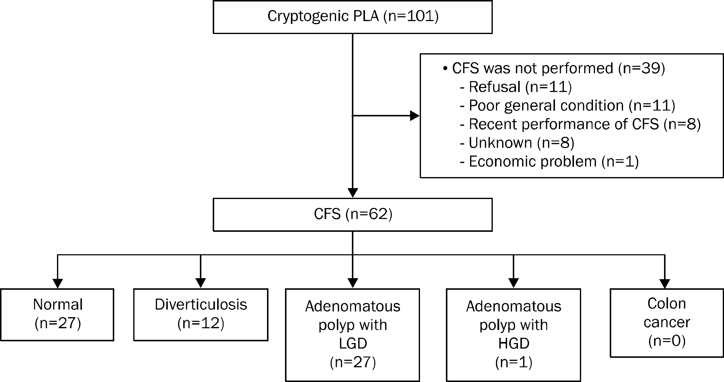
Table 1.
Pyogenic Liver Abscess Etiologies
| Causes | Liver abscess (n=183) |
|---|---|
| Cryptogenic | 101 (55.2) |
| Biliary a | 71 (38.8) |
| Gastrointestinal b | 2 (1.1) |
| Others c | 9 (4.9) |
Table 2.
Baseline Characteristics of Patients with Cryptogenic Pyogenic Liver Abscess
| Characteristic | Cryptogenic liver abscess (n=101) |
|---|---|
| Age (yr) | 62 (20–94) |
| Male | 64 (63.4) |
| White blood cell (/mm3) | 13,190 (1,950–36,480) |
| Hemoglobin (g/dL) | 12.3 (8.4–16.6) |
| Platelet (/mm3) | 170,000 (9,000–759,000) |
| AST (IU/L) | 63 (15–4,336) |
| ALT (IU/L) | 66 (11–1,685) |
| ALP (IU/L) | |
| Hospital 1 a | 331 (120–2,201) |
| Hospital 2, phase I b | 457 (181–2,093) |
| Hosptial 2, phase II b | 172 (37–1,293) |
| GGT (IU/L) | 100 (17–831) |
| Total bilirubin (mg/dL) | 1.0 (0.2–6.6) |
| Albumin (g/dL) | 3.2 (2.0–4.6) |
| Creatinine (mg/dL) | 0.89 (0.40–5.65) |
| Glucose (mg/dL) | 132 (74–503) |
| PT INR | 1.19 (0.95–3.22) |
| CRP (mg/dL) | 17.8 (0.5–40.9) |
| HBsAg c (%) | 6 (6.1) |
| Anti-HCV c (%) | 0 |
| Anti-HIV c (%) | 0 |
| Diabetes mellitus | 38 (37.6) |
| Immune suppressant use | 1 (1.0) |
| Colonoscopic procedure within 1 month | 1 (1.0) |
| Number of abscess pockets | |
| 1 | 75 (74.3) |
| 2 | 18 (17.8) |
| 3 | 4 (4.0) |
| ≥4 | 4 (4.0) |
| Maximal diameter of largest lesion (cm) | 5.7 (1.0–14.0) |
| Percutaneous drainage use | 75 (74.3) |
a Hospital 1 is Inje University Haeundae Paik Hospital, in which the reference range of ALP is 104–338 IU/L.
Table 3.
Esophagogastroduodenoscopic Findings for Cryptogenic Pyogenic Liver Abscess Patients
Table 4.
Causative Bacteria of Cryptogenic Pyogenic Liver Abscess (PLA)
| Bacteria | Cryptogenic PLA (n=101) | PLA with known cause (n=82) |
|---|---|---|
| No growth | 31 (30.7) | 33 (40.2) |
| Klebsiella a | 63 (62.4) | 22 (26.8) |
| Streptococcus | 3 b (2.9) | 6 c (7.3) |
| Escherichia coli | 1 (1.0) | 14 (17.1) |
| Enterobacter | 1 (1.0) | 4 (4.9) |
| Pseudomonas | 1 (1.0) | |
| Gemella morbillorum | 1 (1.0) | |
| Enterococcus | 16 (19.5) | |
| Proteus | 2 (2.4) | |
| Citrobacter | 2 (2.4) | |
| Serratia | 1 (1.2) | |
| Provotella | 1 (1.2) | |
| Shewanella | 1 (1.2) |
 XML Download