

 PDF
PDF ePub
ePub Citation
 Print
Print


Abstract
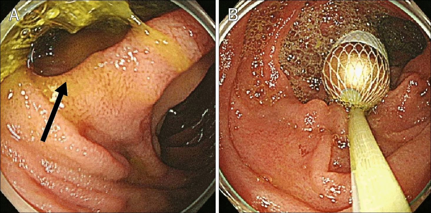
Capsule endoscopy is being increasingly recognized as a gold standard for diagnosing small bowel disease, but along with the increased usage, capsule retention is being reported more frequently. We report a case of capsule endoscopy retention in a diverticulum of the duodenal proximal third portion, which we treated by esophagogastroduodenoscopy. A 69-year-old male visited hospital with hematochezia. He had hypertension and dyslipidemia for several years, and was taking aspirin to prevent heart disease. CT and colonoscopy revealed a diverticulum in the third portion of the duodenum, rectal polyps, and internal hemorrhoids. Capsule endoscopy was performed but capsule impaction occurred. The capsule was later detected by CT in the diverticulum. Endoscopy was performed a day later and the capsule was removed using a net. A small bowel series was conducted after capsule removal, and no stenosis was found. The patient fully recovered and no recurrence of hematochezia was observed at his one month exam. This is the first case in Korea of capsule retention in a duodenal diverticulum, with successful removal by endoscopy.
References
1. Park SK, Ye BD, Kim KO, et al. Korean Gut Image Study Group. Guidelines for video capsule endoscopy: emphasis on Crohn's disease. Clin Endosc. 2015; 48:128–135.

2. Voderholzer WA, Ortner M, Rogalla P, Beinhölzl J, Lochs H. Diagnostic yield of wireless capsule enteroscopy in comparison with computed tomography enteroclysis. Endoscopy. 2003; 35:1009–1014.
3. Liao Z, Gao R, Xu C, Li ZS. Indications and detection, completion, and retention rates of small-bowel capsule endoscopy: a systematic review. Gastrointest Endosc. 2010; 71:280–286.
4. Cave D, Legnani P, de Franchis R, Lewis BS. ICCE. ICCE consensus for capsule retention. Endoscopy. 2005; 37:1065–1067.
5. Cheifetz AS, Lewis BS. Capsule endoscopy retention: is it a complication? J Clin Gastroenterol. 2006; 40:688–691.
6. Courcoutsakis N, Pitiakoudis M, Mimidis K, Vradelis S, Astrinakis E, Prassopoulos P. Capsule retention in a giant Meckel's diverticulum containing multiple enteroliths. Endoscopy. 2011; 43(Suppl 2 UCTN):E308–E309.
7. Ordubadi P, Blaha B, Schmid A, Krampla W, Hinterberger W, Gschwantler M. Capsule endoscopy with retention of the capsule in a duodenal diverticulum. Endoscopy. 2008; 40(Suppl 2):E247–E248.
8. Yang XY, Chen CX, Zhang BL, et al. Diagnostic effect of capsule endoscopy in 31 cases of subacute small bowel obstruction. World J Gastroenterol. 2009; 15:2401–2405.
9. Saperas E, Dot J, Videla S, et al. Capsule endoscopy versus computed tomographic or standard angiography for the diagnosis of obscure gastrointestinal bleeding. Am J Gastroenterol. 2007; 102:731–737.
10. Mylonaki M, Fritscher-Ravens A, Swain P. Wireless capsule endoscopy: a comparison with push enteroscopy in patients with gastroscopy and colonoscopy negative gastrointestinal bleeding. Gut. 2003; 52:1122–1126.
11. Ge ZZ, Hu YB, Xiao SD. Capsule endoscopy and push enteroscopy in the diagnosis of obscure gastrointestinal bleeding. Chin Med J (Engl). 2004; 117:1045–1049.
12. Hara AK, Leighton JA, Sharma VK, Fleischer DE. Small bowel: preliminary comparison of capsule endoscopy with barium study and CT. Radiology. 2004; 230:260–265.
13. Marmo R, Rotondano G, Piscopo R, Bianco MA, Cipolletta L. Meta-analysis: capsule enteroscopy vs. conventional modalities in diagnosis of small bowel diseases. Aliment Pharmacol Ther. 2005; 22:595–604.
14. Ben-Soussan E, Savoye G, Antonietti M, Ramirez S, Lerebours E, Ducrotté P. Factors that affect gastric passage of video capsule. Gastrointest Endosc. 2005; 62:785–790.
Fig. 1.
Initial CT finding. The diverticulum in the proximal 3rd portion of duodenum was detected and its largest diameter was 4.5 cm (arrow).
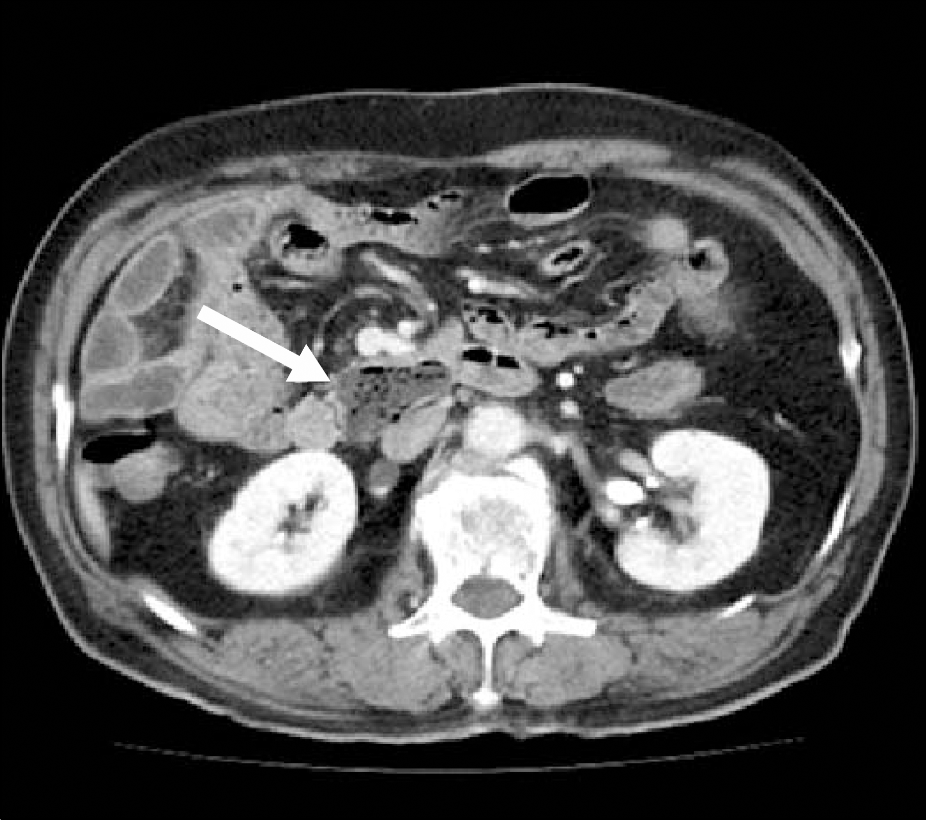
Fig. 2.
Capsule endoscopy findings. The capsule was impacted in the diverticulum, which contained multiple enteroliths at 3 hours after ingestion.
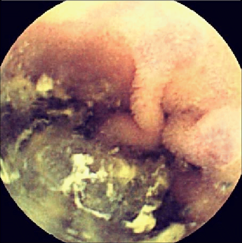
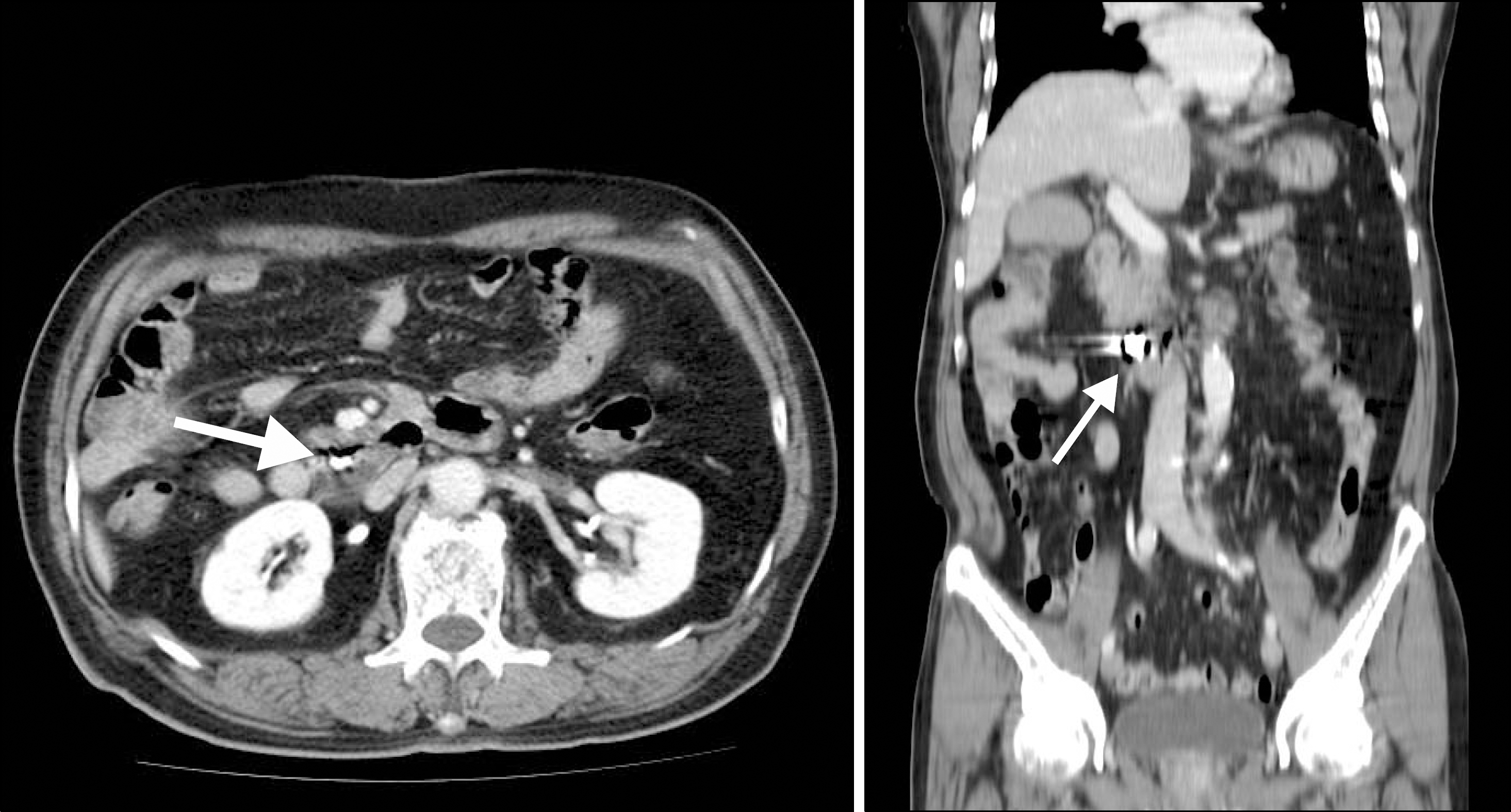
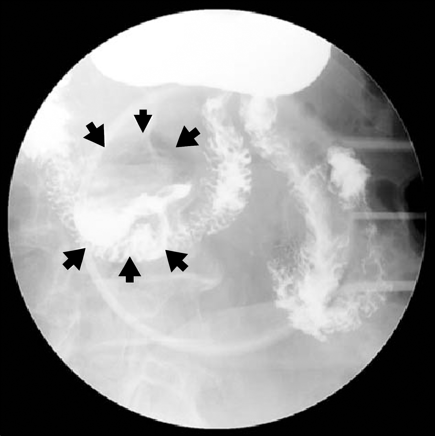
Fig. 3.
Abdominal CT findings. Capsule retention in the diverticulum of duodenal 3rd portion (arrows).
 XML Download