Abstract
A gastric carcinoma with the endoscopic features resembling a submucosal tumor (SMT) is rare, and reportedly accounting for 0.1% to 0.63% of all resected gastric carcinomas in Japan. A diagnosis of a SMT-like gastric carcinoma is often difficult as the tumors are almost entirely covered with normal mucosa. Furthermore mucinous gastric adenocarcinoma is uncommon histologic subtype of gastric cancer. These tumors are detected mostly in an advanced stage and rarely in an early stage. Early mucinous gastric adenocarcinoma is characterized as an elevated lesion resembling SMT due to abundant mucin pools in the submucosa. Here we report one case of SMT-like mucinous gastric adenocarcinoma, diagnosed by the usual endoscopic biopsy and treated with surgery.
References
1. Takemoto N, Baba Y, Kaku Y, et al. Radiologic diagnosis of gastric cancer morphologically mimicking submucosal tumor. Stomach Intest. 1995; 30:759–768.
2. Umehara Y, Kimura T, Okubo T, et al. Gastric carcinoma resembling submucosal tumor. Gastric Cancer. 1999; 2:191–193.

3. Ryu CB, Cho JY, Lee JS, Lee MS, Jin SY, Shim CS. Mucinous gastric adenocarcinoma with morphological change from polypoid to depressed lesion within a short period. Endoscopy. 2002; 34:1026.

4. Watanabe H, Enjoji M, Imai T. Gastric carcinoma with lymphoid stroma. Its morphologic characteristics and prognostic correlations. Cancer. 1976; 38:232–243.
5. Park SW, Jo YJ, Lee JY, et al. A case of mucinous gastric adenocarcinoma as submucosal tumor. Korean J Gastroenterol. 2004; 44:47–49.
6. Ohara N, Tominaga O, Uchiyama M, Nakano H. A case of advanced gastric cancer resembling submucosal tumor of the stomach. Jpn J Clin Oncol. 1997; 27:423–426.

7. Chonan A, Mochizuki F, Fujita N, et al. Endoscopic diagnosis of gastric cancer similar to submucosal tumors. Stomach Intest. 1995; 30:777–785.
8. Nishinaka H, Kodama K, Iwamura M, et al. Gastric cancer derived from heterotopic gastric glands, report of a case. Stomach Intest. 2003; 38:1250–1254.
9. Japanese Gastric Cancer Association. Japanese classification of gastric carcinoma. 2nd English edition. Gastric Cancer. 1998; 1:10–24.
10. Yuki T, Sato T, Ishida K, et al. Clinicopathological and imaging features of gastric carcinoma resembling submucosal tumor. Stomach Intest. 2003; 38:1215–1224.
11. Nam JH, Park SJ, Park JE, et al. Two cases of submucosal tumorlike gastric adenocarcinoma. Korean J Gastrointest Endosc. 2007; 34:94–98.
12. Kim SY, Park JJ, Cho Y, et al. A case of submucosal tumorlike early gastric adenocarcinoma diagnosed by endoscopic mucosal resection. Korean J Gastrointest Endosc. 2005; 31:404–408.
13. Uedo N, Iishi H, Ishiguro S, et al. Mucinous adenocarcinoma of the stomach mimicking submucosal tumor, report of a case. Stomach Intest. 2003; 38:1245–1249.
14. Takahashi T, Otani Y, Yoshida M, et al. Gastric cancer mimicking a submucosal tumor diagnosed by laparoscopic excision biopsy. J Laparoendosc Adv Surg Tech A. 2005; 15:51–56.

15. Nakamura T, Suzuki T, Kobayshi S, et al. Diagnosis of gastric carcinoma with the appearance of a submucosal tumor of the stomach, using endoscopic ultrasonography. Stomach Intest. 1995; 30:787–798.
16. Endo T, Okuda H, Arimura Y, et al. A case of early gastric carcinoma with lymphoid stroma: diagnostic usefulness of endo-sonography. Dig Endosc. 1998; 10:240–243.

17. Fujiyoshi A, Kawamura M, Ishitsuka S. Gastric adenocarcinoma mimicking a submucosal tumor: case report. Gastrointest Endosc. 2003; 58:633–635.
18. Yasuda K, Shiraishi N, Inomata M, Shiroshita H, Ishikawa K, Kitano S. Clinicopathologic characteristics of early-stage mucinous gastric carcinoma. J Clin Gastroenterol. 2004; 38:507–511.

19. Jo MA, Kim SH, Kim SH, et al. A case of gastritis cystica profunda with early gastric cancer. Korean J Med. 2004; 67:78–82.
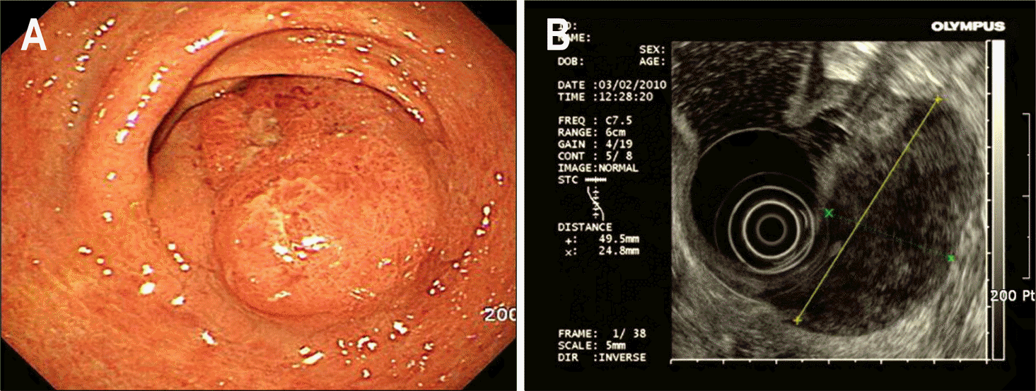
Fig. 1.
Gastroscopic and endoscopic ultrasonographic findings. (A) They showed elevated lesion with focal erosion and ulcer in the greater curvature of the gastric lower body to antrum. (B) Reticular high echogenic speckeles were observed in the low echogenic tumor.

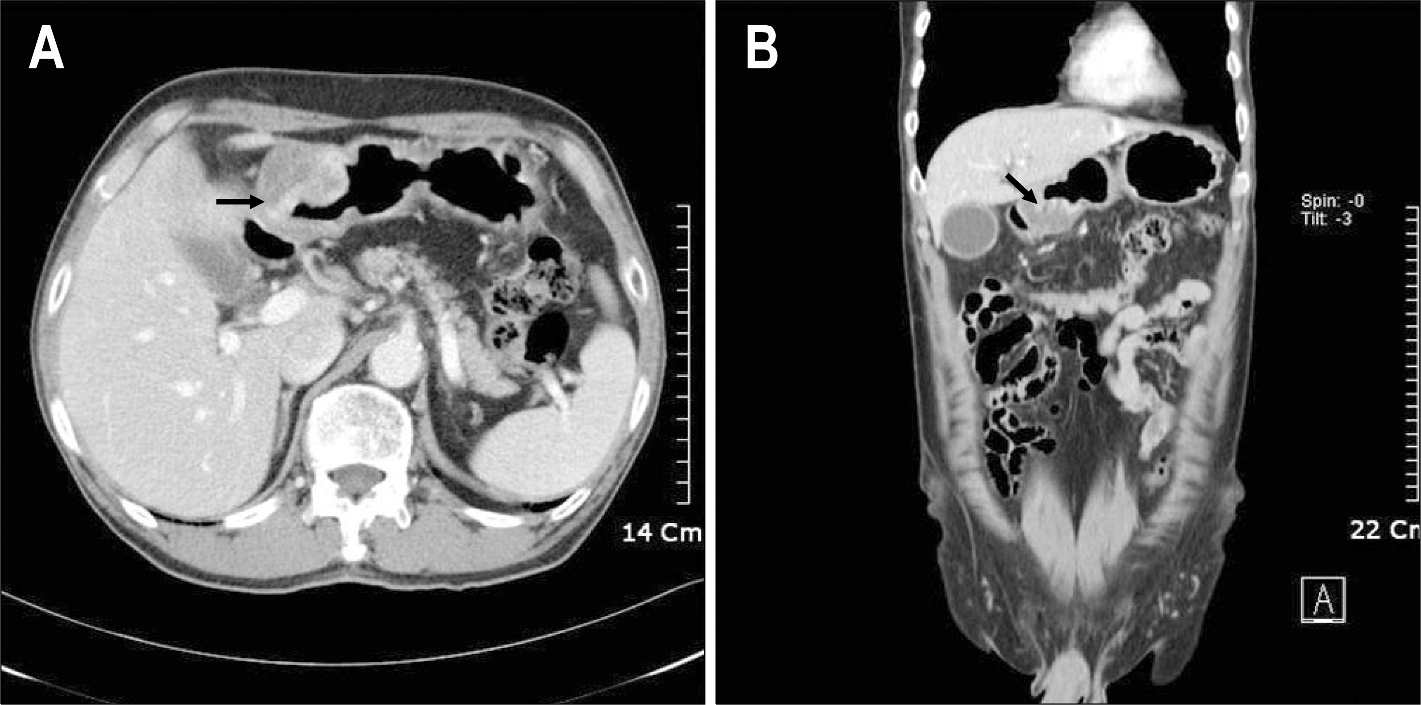
Fig. 2.
Abdominal CT findings. They showed well enhanced mass lesion (arrow) with inhomogenous nature in the greater curvature of the gastric lower body to antrum.

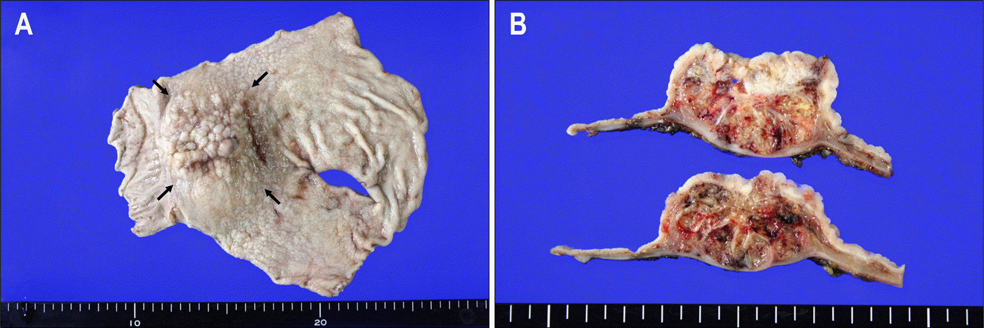
Fig. 3.
Gross findings of the resected stomach. (A) A round bulged-out mass (arrow) was located on the antrum, measuring about 6×5.5 cm in size. The mucosal surface showed nodularity and focal superficial ulceration. (B) On cross section, the tumor infiltrated into the proper muscle and subserosa. It was mostly composed of gelatinous mucin and focal whitish tan thickened mucosal lesion with relatively normal appearing overlying mucosa.


 PDF
PDF ePub
ePub Citation
 Print
Print


 XML Download