

 PDF
PDF ePub
ePub Citation
 Print
Print


INTRODUCTION
Nuts are rich in nutrients and bioactive food components that exhibit anti-cancer properties. Recent prospective studies have suggested the protective effects of nut consumption on colorectal cancer [1]. Walnuts (Juglans regia L.) are well known to have many health effects such as anti-inflammatory and anti-oxidant properties as well as cardioprotective effect [234]. Walnuts, in particular, contain multiple components such as omega-3 fatty acids, phytochemicals, fiber, phenolic compounds, folate, and vitamin E, each of which has been individually associated with reducing the risk of certain cancers. In a mouse study, a walnut-containing diet was shown to inhibit colorectal cancer growth by suppressing angiogenesis; previous observations also demonstrated walnut extracts to have inhibitory effects on the growth of colon cancer cells [5]. Moreover, we have already demonstrated that walnut phenolic extracts (WPEs) can suppress the growth of colon cancer stem cells [6].
Telomeres, located at the chromosomal ends and getting shorter during each cell cycle, are crucial for chromosomal integrity and stability. Shortened telomere length has been associated with risks of cancers and age-associated diseases. In fact, maintenance of telomere length is essential for cell replication and tumorigenesis and telomere shortening is an initial event in colorectal carcinogenesis [78].
Telomerase, an RNA-dependent DNA polymerase that synthesizes telomeric DNA sequences, is induced for cell proliferation and inhibited for cell differentiation, indicating that the regulation of telomerase activity could be a mechanism that controls the cell growth [9]. Thus, telomerase can be a target for cancer therapy. In fact, telomerase activity is present in over 90% of cancerous cells and immortalized cells but absent in most normal human somatic cells. Downregulation of telomerase activity is associated with cell cycle arrest and apoptosis in colon cancer [10].
The human telomerase reverse transcriptase (hTERT), a catalytic subunit of the telomerase, is upregulated in the majority of immortalized cells and tumor cells but not expressed in most primary somatic human cells and tissues. In fact, hTERT transcription is strictly controlled and closely associated with telomerase activity, suggesting that hTERT is the upstream gene to control the telomerase enzyme activity [11]. Interestingly, hTERT is a target of c-MYC that has several binding sites on hTERT promoter and stimulates hTERT promoter activity through its overexpression. It is also well known that c-MYC is a powerful proto-oncogene that controls many facets of cell growth and metabolism [12].
To investigate the mechanism behind the inhibitory effect of walnuts on colon cancer cell growth we determined the telomere length and telomerase activity using a colon cancer stem cell model that we have used in the previous studies [613]. We also measured the transcription levels of hTERT and c-MYC to determine the upstream effect of WPE on telomere maintenance.
MATERIALS AND METHODS
Preparation of WPE and cell culture
WPE was prepared using English walnuts (Juglans regia L.) obtained from the California Walnut Commission by performing methanolic extraction, as described previously [14]. The human colon cancer cell line HCT116, purchased from ATCC (Rockville, MD, USA), was cultured in McCoy's 5A medium (WelGENE, Daegu, Korea) with 10% (v/v) fetal bovine serum (HyClone, Logan, UT, USA) and 1% (v/v) penicillin-streptomycin (Invitrogen, Carlsbad, CA, USA). The cells were analyzed using the FACSDiVa system (BD, San Jose, CA, USA) to detect the expression of the CSC markers, CD133 and CD44, with Alexa Fluor 488-conjugated anti-CD133 monoclonal antibody (Miltenyi Biotec, Bergisch Gladbach, Germany) and anti-CD44 monoclonal antibody (Cell Signaling, Danvers, MA, USA), respectively, as described previously [15]. CD133+CD44+ HCT116 cells were treated with WPE at the concentrations of 0, 10, 20, and 40 µg/mL in triplicate for 6 days.
Telomere length
Telomere lengths were assessed by quantitative real-time PCR using telomere specific primers and genomic DNA extracted from the cells. The telomere length was represented by the ratio of the telomere repeat copy number (T) to the single-copy gene copy number (S), which was further adjusted with the T/S ratio of a reference DNA sample. The 36B4 gene located on chromosome 12q24 was used for the single-copy number gene and the human embryonic kidney 293 (HEK 293) cell line was used as the reference DNA. Genomic DNA was isolated using a DNA extraction kit (Qiagen, Hilden, Germany) via the spin-column method. The reference DNA was sequentially three-fold diluted into 5 different template amounts ranging from 0.21 ng to 16.67 ng and the standard curve was constructed by plotting the each Ct value. qRT-PCR was performed in triplicate with each primer as described previously [16]. The primer sequences are in Table 1. The total volume of the PCR mixture was 20 µL; 10 µL of 1 × SYBR qPCR master mix (Takara Shuzo, Kyoto, Japan), 2 µL of 20 ng template, 0.2 µL of 1× ROX dye, 2 uL of each primer, and the rest volume with nuclease free water. Primers were added to final concentrations of 100 nmol/L for the telomere forward primer, 900 nmol/L for the telomere reverse primer, 300 nmol/L for the single-copy gene forward primer, and 500 nmol/L for the single-copy gene reverse primer. For the telomeric qRT-PCR, polymerase enzymes were activated at 95℃ for 10 min, followed by 35 cycles of 95℃ for 30 sec and 57.5℃ for 30 sec. The single-copy gene PCR reaction consisted of 40 cycles of 95℃ for 30 sec and 57.5℃ for 30 sec after a 95℃ for 10 min cycle using Applied Biosystems Viia7 (Foster City, CA, USA). The amplification specificity was determined via melting curve analysis. The T/S ratio (dCt) for each sample was calculated by subtracting the Ct value of the single-copy gene from the Ct value of each telomere PCR reaction. The relative T/S ratio (ddCt) was adjusted with respect to the Ct of control HEK 293 cells.
Telomerase activity
Telomerase activity was measured via the PCR-based method using lysed cells. The PCR reaction mixture consisted of SYBR Green master mix (Takara Shuzo, Kyoto, Japan), ethylene glycol tetra acetic acid (EGTA), forward primer (TS), reverse primer (ACX), and protein extract from the cells. Primer sequences are shown in Table 1 [17]. The PCR reaction mixture was incubated at room temperature for 30 min to allow the telomerase in the protein extracts to elongate the TS primer by adding TTAGGG repeat sequences. Thereafter, qRT-PCR was conducted with the following PCR condition: 95℃ incubation for 10 min followed by 40 cycles of 95℃ for 20 sec and 50℃ for 30 sec. All samples were run in triplicate. The HEK 293 cell line was used as a reference and telomerase activity (Mean±SE) was represented as dCt.
hTERT and c-MYC transcriptions
Quantitation of mRNA expression was performed by reverse transcription polymerase chain reaction (RT-PCR). Total RNA was extracted using Nucleozol (MACHEREY-NAGEL GmbH & Co. KG, Postfach, Düren, Germany) from CD133+CD44+ HCT116 cells treated with various concentrations of WPE. The RNA was transcribed into cDNA using AccuPower® RT PreMix (Bioneer, Daejeon, Korea) and amplified using primers specific to each gene and PCR master mix (Bioneer, Daejeon, Korea). The PCR products were separated by 1.5% agarose gel electrophoresis. Each mRNA level was normalized with Ribosomal protein large subunit P 0 (RPLP0). All primers are shown in Table 1 [1819202122].
Statistical analysis
Differences between the variables of the telomere length and telomerase activity at each concentration of WPE were tested using ANOVA. Duncan's post hoc analysis using variance was also performed to compare the differences between the groups. Correlations between telomere length and telomerase activity were assessed using Spearman's correlation. P<0.05 was regarded as significant. All statistical analyses were performed using SPSS version 24.0 (IBM Corporation, Armonk, NY, USA).
RESULTS
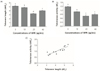
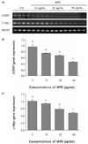
Telomere length in WPE-treated cells was significantly decreased in a dose-dependent manner up to the 40 µg/mL concentration (5.16 ± 0.13 at 0 µg/mL, 4.79 ± 0.12 at 10 µg/mL, 3.24 ± 0.08 at 20 µg/mL, and 3.99 ± 0.09 at 40 µg/mL; P = 0.0276) (Fig. 1A). At 40 µg/mL, telomere length was still significantly lower than the control, but higher compared to the 20 µg/mL concentration. Interestingly, telomerase activities concurrently decreased with telomere length up to the 40 µg/mL concentration (1.47 ± 0.04, 1.09 ± 0.01, 0.76 ± 0.08, and 0.88 ± 0.06; P = 0.0067) (Fig. 1B), even though no additive effect was observed at 40 µg/mL compared to 20 µg/mL. Significant positive correlation was also observed between the telomere length and telomerase activity (Fig. 1C; r = 0.9090, P < 0.0001). The transcriptions of hTERT showed a significant stepwise decreasing pattern by stepwise increasing WPE concentrations (Fig. 2A; P < 0.0001). The transcription of c-MYC, which is known to involve in the transcription of hTERT, also showed a significant stepwise decreasing pattern by WPE (Fig. 2B; P < 0.0001). Collectively, WPE treatment simultaneously reduced the transcriptions of c-MYC and hTERT as well as telomerase activity and telomere length.
DISCUSSION
Telomere maintenance is a prerequisite for indefinite proliferation in cancer. Therefore, inhibiting the maintenance of telomeres may prove to be a useful strategy for cancer therapy. The telomere and telomerase connection can obviously offer several targets for inducing cancer cell death [23]. In the present study, we demonstrated that WPE reduced telomerase activity and telomere length as well as transcriptions of hTERT and c-MYC, both of which are known to control telomerase activity. It could be a mechanism by which walnuts inhibit the growth of colon cancer cells [6]. To the best of our knowledge, this is the first study that attempts to determine the effect of walnuts on telomere maintenance.
Telomere length, which is crucial for individual cell senescence, is also highly associated with cancer. Individuals with short telomeres are known to have an increased risk of cancer because short telomeres lead to genomic instability; however, individuals with long telomeres paradoxically can have an increased risk of major cancers [24]. The association between the blood cell telomere length and cancer risk is also supported by a meta-analysis which demonstrated that ever smokers have shorter telomeres compared with never smokers. Furthermore, current smokers have shorter telomeres compared with never or former smokers [25]. In a systemic review that analyzed the association between tissue telomere length and breast cancer prognosis, longer telomeres were associated with better prognosis and shorter telomeres were associated with higher local recurrence rates and higher tumor grade [26]. Collectively, telomere length can be a good biomarker for the development and prognosis of cancer, suggesting that telomere and telomere metabolism can be a potential target for cancer treatment. We, therefore, investigated the effects of walnut on telomere length and telomerase activity as well as the expression of upstream gens using a colon cancer stem cell model in which walnut have shown to reduce the cell proliferation and viability [6].
Telomerase-targeted treatments for cancer have garnered a lot of attention because of the peculiar characteristics of telomerases, which are detected in nearly all cancer cells but not expressed in most normal cells [27]. Numerous studies have shown that the inhibition of telomerase induces cancer cell death. Sulforaphane, a bioactive food component found abundantly in broccoli and broccoli sprouts, can inhibit cell viability and induce apoptosis, simultaneously downregulating the protein expression and enzymatic activity of telomerase in colon cancer cell lines owing to its epigenetic effect as a histone deacetylase [28]. In a previous study, a certain form of L-asparaginases, which were used to treat acute lymphoblastic leukemia with some limitations, was found to suppress telomerase activity in human T-cell lymphoma Jurkat cells; this is considered to be an additional anticancer mechanism of these compounds, and inhibition of the telomerase activity may be considered to be a promising approach for cancer treatment [29]. It appears that the decreased telomerase activity demonstrated in this study can be a mechanism by which WPE reduces telomere length and thereby reduces cell viability. Interestingly, the positive correlation between telomere length and telomerase activity further supports this mechanism. A growing body of evidence has also demonstrated that telomere stability can be affected more than the main genome by conventional cancer chemotherapeutic agents, suggesting that cancer chemotherapies with genotoxic properties preferably target telomere [30].
Collectively, our observation that WPE reduced telomerase activity and telomere length represents the potential effect to assist cancer chemotherapy.
We also found that the repression of hTERT and c-MYC by WPE was in parallel with decreasing telomerase length and telomerase activity, suggesting that the effect of WPE on telomere maintenance could be conveyed through those genes. Because hTERT is a catalytic subunit of the enzyme telomerase, it is not surprising that the hTERT expression was downregulated by WPE simultaneously with the reduction of telomerase activity. On the other hand, the observation that WPE reduced the expression of c-MYC, which can affect the expression of hTERT, is quite interesting, because c-MYC plays many other important roles in cancer including the transformation of normal human cells into cancer cells [11]. The c-MYC transcription factor is involved in cellular growth, proliferation and metabolism by controlling the expression of numerous target genes [31]. It suggests that walnut may have another anticancer property through the inhibition of c-MYC transcription.
In conclusion, WPE demonstrates the ability to disrupt telomere maintenance in a colon cancer stem cells model. Further studies are warranted to determine the cause and effective relationship between the inhibition of telomere maintenance by WPE and the colon cancer stem cell viability.

 XML Download